. 2023 Jul 13;15(7):1539.
doi: 10.3390/v15071539.

Analogs of the Catechol Derivative Dynasore Inhibit HIV-1 Ribonuclease H, SARS-CoV-2 nsp14 Exoribonuclease, and Virus Replication

Affiliations

Analogs of the Catechol Derivative Dynasore Inhibit HIV-1 Ribonuclease H, SARS-CoV-2 nsp14 Exoribonuclease, and Virus Replication

Abhishek Asthana et al. Viruses. .

Abstract

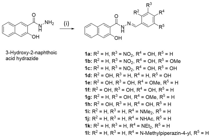
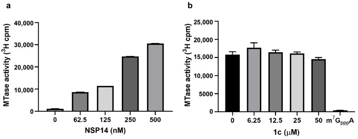
Viral replication often depends on RNA maturation and degradation processes catalyzed by viral ribonucleases, which are therefore candidate targets for antiviral drugs. Here, we synthesized and studied the antiviral properties of a novel nitrocatechol compound (1c) and other analogs that are structurally related to the catechol derivative dynasore. Interestingly, compound 1c strongly inhibited two DEDD box viral ribonucleases, HIV-1 RNase H and SARS-CoV-2 nsp14 3'-to-5' exoribonuclease (ExoN). While 1c inhibited SARS-CoV-2 ExoN activity, it did not interfere with the mRNA methyltransferase activity of nsp14. In silico molecular docking placed compound 1c in the catalytic pocket of the ExoN domain of nsp14. Finally, 1c inhibited SARS-CoV-2 replication but had no toxicity to human lung adenocarcinoma cells. Given its simple chemical synthesis from easily available starting materials, these results suggest that 1c might be a lead compound for the design of new antiviral compounds that target coronavirus nsp14 ExoN and other viral ribonucleases.

Keywords: COVID-19; HIV; HIV-1 RNase H; SARS-CoV-2 nsp14; antivirals; coronavirus; enzyme inhibitors; ribonucleases.


Conflict of interest statement

The authors declare no conflict of interest.

Figures

Scheme 1
Scheme 1
Reagents and conditions: (i) Aryl aldehyde, EtOH, reflux, 2 h, 49–87%.
Scheme 2
Scheme 2
Reagents and conditions: (i) Acid hydrazide, EtOH, reflux, 2 h, 64–86%.
Figure 1
Figure 1
Inhibition of ExoN activity of nsp14/10 complex by 1c and its analogs. ExoN activity of SARS-CoV-2 nsp14/nsp10 was determined at different concentrations of inhibitors in an in vitro assay using HPLC to monitor degradation of the RNA22 substrate. The inhibitors used were 1c and its analogs 2a, 2b, and 2c. An IC50 of 2.2 µM was calculated for inhibitor 1c. Inhibition of ExoN activity by the analogs is shown at 100 µM concentration, as indicated on the graph. RNA22 substrate incubated under the same conditions (in the absence of both nsp14/10 and inhibitors) served as un-degraded control (100% intact substrate). Experiments were performed in triplicate. Error bars show standard deviations.
Figure 2
Figure 2
Effect of inhibitor 1c on MTase activity of full-length SARS-CoV-2 nsp14. (a) Increasing concentrations of nsp14 were incubated with GpppA for 60 min in the presence of [3H]-SAM in a radioactive biochemical assay. (b) Nsp14 MTase activity monitored in the absence or presence of inhibitor 1c using [3H]-SAM. m7GpppA served as a methylated control substrate, performed in the absence of 1c. Experiments were performed in triplicate. Error bars show standard deviations.
Figure 3
Figure 3
Here, 1c binds in the exonuclease catalytic site of the nsp14-nsp10 complex. In silico docking simulation predicted specific binding of compound 1c in the exonuclease catalytic site. (a) Electrostatic surface potential of nsp14-nsp10 complex with dsRNA substrate. (b) The binding pocket view of 1c in the ExoN catalytic site. Negative (red) and positive (blue) surface potentials are shown. (c) Side chains of the nsp14 amino acid within 5 Å of compound 1c in the ExoN catalytic site. Residues are depicted as sticks, while 1c is depicted using ball-and-stick modeling with elemental color representations. Mg2+ ions are shown in green and the dotted line shows the possible interaction with Mg2+. Amino acid residues, as sticks, and inhibitor 1c, in ball and sticks, are depicted using elemental colors.
Figure 4
Figure 4
Compound 1c was not toxic to A549 cells. A549 cells were treated with varying concentrations of 1c (0.75 to 50 µM) for 24, 48, 72, or 96 h. Percent of live cells were determined and compared to DMSO (v/v) controls using an Alamar-Blue-based fluorescence (Ex/Em 560/590 nm) assay. The experiments were performed in triplicate. Error bars represent standard deviations.
Figure 5
Figure 5
SARS-CoV-2 replication inhibition by 1c in A549 ACE2+ cells. A549 ACE2+ cells were infected at an MOI of 0.05 and treated with varying concentrations of 1c (12.5, 25, 50 µM) for 24 or 48 h. The experiments were performed twice in triplicate (N = 6). Statistical significance was calculated using unpaired t-test (ns, not significant; ****, p < 0.0001). Error bars represent standard deviations.
Figure 6
Figure 6
Plaque reduction assay. (a) SARS-CoV-2 replication inhibition by 1c and 2ac at the indicated concentrations for 72 h. Statistical significance was calculated using unpaired t-test (**, p < 0.01; ***, p < 0.001; ****, p < 0.0001). (b) Representative images of wells in a plaque reduction assay are shown. Inhibitors and their concentrations are indicated on the panels.

References

    1. Chen B., Tian E.-K., Tian L., Han R., Wang S., Xiang Q., Zhang S., El Arnaout T., Cheng W. Overview of lethal human coronaviruses. Signal Transduct. Target. Ther. 2020;5:89. doi: 10.1038/s41392-020-0190-2. - DOI - PMC - PubMed
    1. Hu B., Guo H., Zhou P., Shi Z.-L. Characteristics of SARS-CoV-2 and COVID-19. Nat. Rev. Microbiol. 2021;19:141–154. doi: 10.1038/s41579-020-00459-7. - DOI - PMC - PubMed
    1. Markov P.V., Ghafari M., Beer M., Lythgoe K., Simmonds P., Stilianakis N.I., Katzourakis A. The evolution of SARS-CoV-2. Nat. Rev. Microbiol. 2023;21:361–379. doi: 10.1038/s41579-023-00878-2. - DOI - PubMed
    1. Singh D., Yi S.V. On the origin and evolution of SARS-CoV-2. Exp. Mol. Med. 2021;53:537–547. doi: 10.1038/s12276-021-00604-z. - DOI - PMC - PubMed
    1. Al-Awwal N., Dweik F., Mahdi S., El-Dweik M., Anderson S.H. A review of SARS-CoV-2 disease (COVID-19): Pandemic in our time. Pathogens. 2022;11:368. doi: 10.3390/pathogens11030368. - DOI - PMC - PubMed

Publication types