

Left atrial sphericity in relation to atrial strain and strain rate in atrial fibrillation patients
- PMID: 37515682
- PMCID: PMC10520187
- DOI: 10.1007/s10554-023-02866-2
Left atrial sphericity in relation to atrial strain and strain rate in atrial fibrillation patients
Abstract
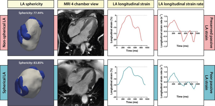
Purpose: Left atrial (LA) sphericity is a novel, geometry-based parameter that has been used to visualize and quantify LA geometrical remodeling in patients with atrial fibrillation (AF). This study examined the association between LA sphericity, and LA longitudinal strain and strain rate measured by feature-tracking in AF patients.
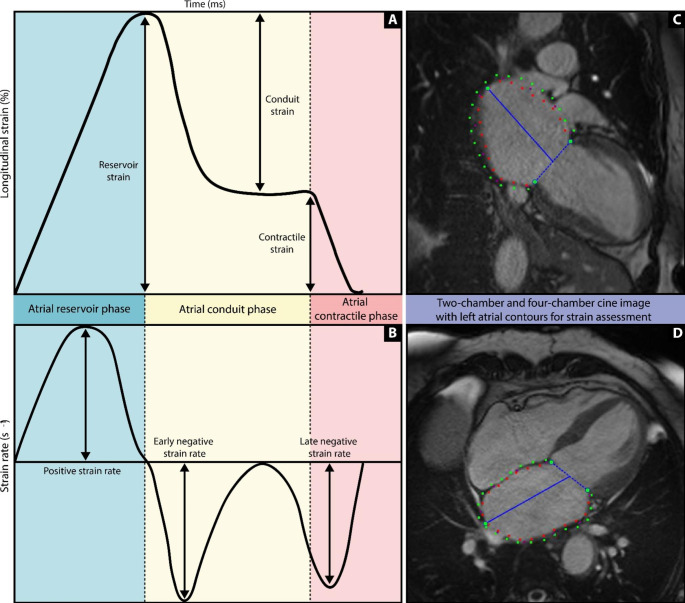
Methods: 128 AF patients who underwent cardiovascular magnetic resonance (CMR) imaging in sinus rhythm prior to their pulmonary vein isolation (PVI) procedure were retrospectively analyzed. LA sphericity was calculated by segmenting the LA (excluding the pulmonary veins and the LA appendage) on a 3D contrast enhanced MR angiogram and comparing the resulting shape with a perfect sphere. LA global reservoir strain, conduit strain, contractile strain and corresponding strain rates were derived from cine images using feature-tracking. For statistical analysis, Pearson correlations, multivariable logistic regression analysis, and Student t-tests were used.
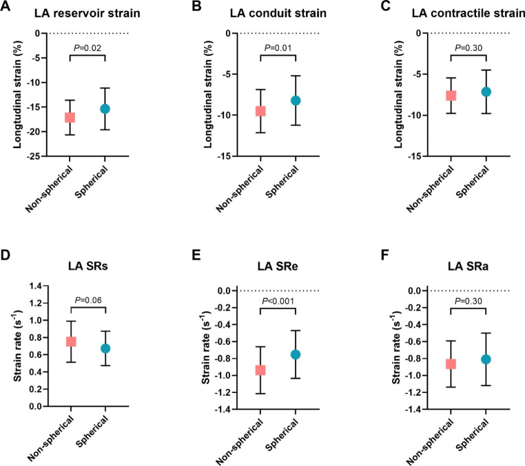
Results: Patients with a spherical LA (dichotomized by the median value) had a lower reservoir strain and conduit strain compared to patients with a non-spherical LA (-15.4 ± 4.2% vs. -17.1 ± 3.5%, P = 0.02 and - 8.2 ± 3.0% vs. -9.5 ± 2.6%, P = 0.01, respectively). LA strain rate during early ventricular diastole was also different between both groups (-0.7 ± 0.3s- 1 vs. -0.9 ± 0.3s- 1, P = 0.001). In contrast, no difference was found for LA contractile strain (-7.2 ± 2.6% vs. -7.6 ± 2.2%, P = 0.30).
Conclusions: LA passive strain is significantly impaired in AF patients with a spherical LA, though this relation was not independent from LA volume.
Keywords: Atrial fibrillation; Atrial remodeling; Atrial sphericity; Atrial strain; Cardiac MRI.
© 2023. The Author(s).
Conflict of interest statement
Authors have nothing to disclose.
Figures


References
-
- Benito EM, Carlosena-Remirez A, Guasch E, Prat-González S, Perea RJ, Figueras R, et al. Left atrial fibrosis quantification by late gadolinium-enhanced magnetic resonance: a new method to standardize the thresholds for reproducibility. Europace. 2016;19(8):1272–1279. doi: 10.1093/europace/euw219. - DOI - PubMed
-
- Nakamori S, Ngo LH, Tugal D, Manning WJ, Nezafat R. Incremental Value of Left Atrial Geometric Remodeling in Predicting Late Atrial Fibrillation Recurrence After Pulmonary Vein Isolation: A Cardiovascular Magnetic Resonance Study. Journal of the American Heart Association. 2018;7(19):e009793. - PMC - PubMed
-
- Gloschat C, Cates J, Walker B, MacLeod RS. Statistical shape modeling of the left atrium from MRI of patients with atrial fibrillation. J Cardiovasc Magn Reson. 2011;13(1):P57. doi: 10.1186/1532-429X-13-S1-P57. - DOI
MeSH terms
LinkOut - more resources
Full Text Sources
Medical
