. 2023 Oct 2;220(10):e20220953.
doi: 10.1084/jem.20220953. Epub 2023 Jul 26.

Skin mesenchymal niches maintain and protect AML-initiating stem cells

Affiliations

Skin mesenchymal niches maintain and protect AML-initiating stem cells

Lakshmi Sandhow et al. J Exp Med. .

Abstract

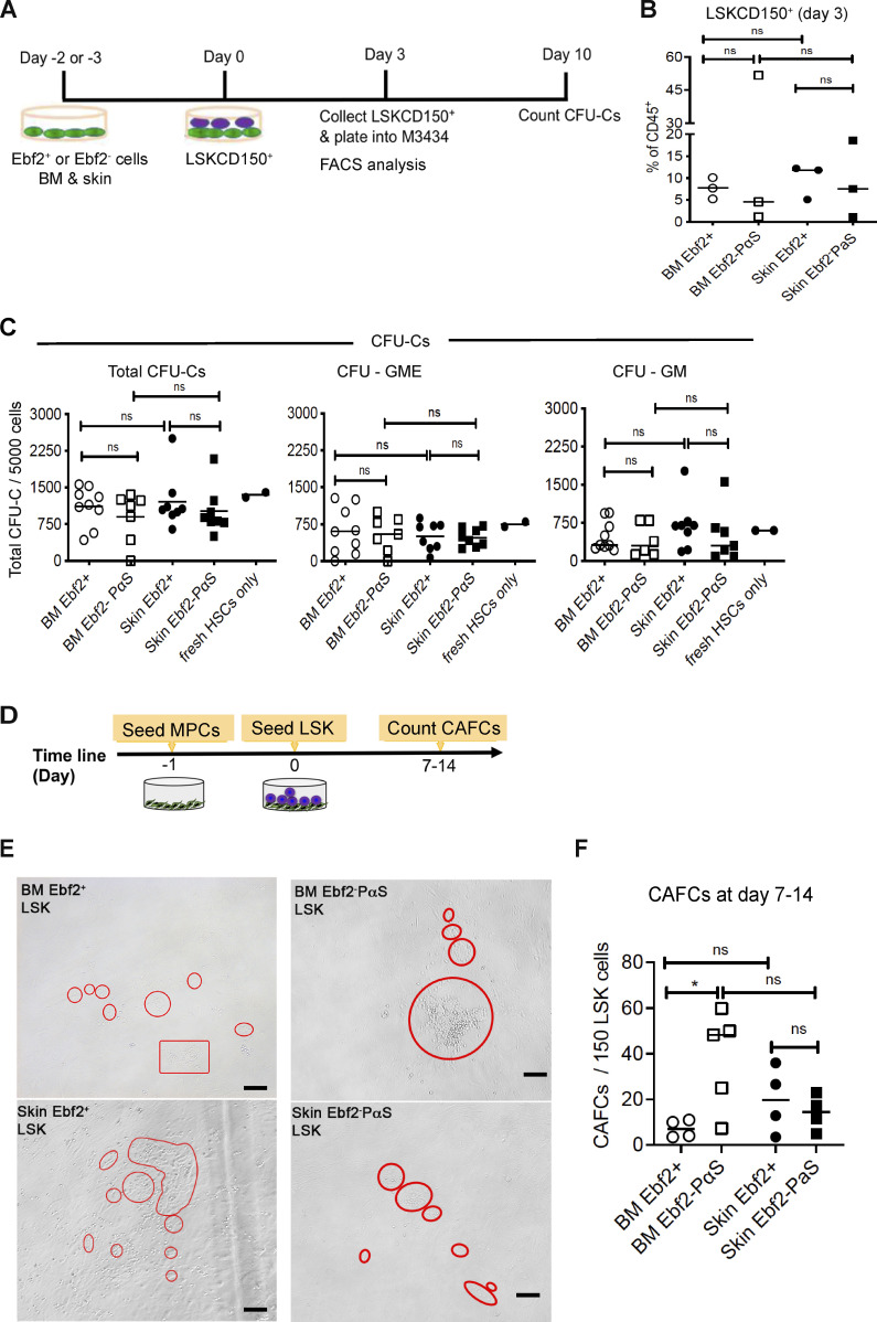
Leukemia cutis or leukemic cell infiltration in skin is one of the common extramedullary manifestations of acute myeloid leukemia (AML) and signifies a poorer prognosis. However, its pathogenesis and maintenance remain understudied. Here, we report massive AML cell infiltration in the skin in a transplantation-induced MLL-AF9 AML mouse model. These AML cells could regenerate AML after transplantation. Prospective niche characterization revealed that skin harbored mesenchymal progenitor cells (MPCs) with a similar phenotype as BM mesenchymal stem cells. These skin MPCs protected AML-initiating stem cells (LSCs) from chemotherapy in vitro partially via mitochondrial transfer. Furthermore, Lama4 deletion in skin MPCs promoted AML LSC proliferation and chemoresistance. Importantly, more chemoresistant AML LSCs appeared to be retained in Lama4-/- mouse skin after cytarabine treatment. Our study reveals the characteristics and previously unrecognized roles of skin mesenchymal niches in maintaining and protecting AML LSCs during chemotherapy, meriting future exploration of their impact on AML relapse.


Conflict of interest statement

Disclosures: The authors declare no competing financial interests.

Figures

Figure 1.
Figure 1.
AML cells infiltrated in skin are capable of regenerating AML after transplantation. (A) Strategy to generate MLL-AF9 AML mouse model. MLL-AF9 transduced AML cells expressing CD45.1 were transplanted into non-irradiated CD45.2 C57BL/6 mice. (B) FACS profile showing AML engraftment analysis in BM, PB, spleen, and dorsal skin at the end stage of AML. The numbers in the panel are the mean frequencies. (C) Proportion of the AML cells within total hematopoietic (CD45+) cells in blood, skin, BM, and spleen. Data were from two independent experiments and each dot represents data from one mouse. The horizontal bars represent mean values. n = 4–9 per group, *P < 0.05, **P < 0.01, ****P < 0.0001, determined by paired t test, except the comparison between skin and blood where unequal number of mice were included. (D) The inverse correlation between AML engraftment in the skin with symptomatic AML onset. *P < 0.05, determined by Spearman correlation. Each dot represents data from a single recipient mouse, n = 18 mice. (E) Confocal image showing distribution of MLL-AF9+ AML cells expressing DsRed at perivascular sites in dorsal skin. The AML cells were injected after sublethal irradiation of the mice. The endothelial cells were marked by CD31 expression (white). Scale bars are 50 µm. (F) Experimental strategy to assess AML-initiating capacity of the AML cells infiltrated in mouse skin by serial transplantation. The primary recipient mice (CD45.2) that developed AML after AML cell (CD45.1) transplantation were treated with Ara-C (100 mg/Kg) or saline (NS, untreated) for 5 d. The residual CD45.1+ AML cells from skin were sorted at 2–3 d after the last injection of Ara-C and transplanted into secondary non-irradiated recipient CD45.2 mice at doses of 10, 100, 1,000, and 5,000 (5K) cells/mouse. The AML development was monitored by FACS and hematology analyzer Sysmex. The mice were sacrificed when found moribund. (G) AML engraftment in blood, skin, BM, and spleen of the primary recipients under steady state and 3 d after Ara-C treatment. Each dot represents data from a single mouse. n = 5 per group, *P < 0.05, ***P < 0.001, determined by paired t test. (H) The fraction of CD36+ AML cells in the skin, BM, and spleen. Each dot represents data from a single mouse. ns, no significant difference, determined by paired t test. (I) The frequency of CFU-Cs derived from the residual AML cells in skin, spleen, and BM. (J) The Kaplan–Meier survival curve of the primary and secondary recipient mice. The curve was generated by Log-rank (Mantel–Cox test). Each dot represents an endpoint when the mice were found dead or sacrificed because of being moribund. Data were from two independent experiments. n = 5–8 primary recipients treated with Ara-C (n = 5) or NS (untreated), n = 5–7 secondary recipients per group. (K) Platelet counts in PB of the secondary recipient mice at the endpoint. Each dot represents data from a single mouse. (L) Spleen size of the secondary recipient mice at the endpoint. Each dot represents data from a single mouse. (M) FACS profile showing the engraftment of skin-derived AML CD45.1+ cells in BM of the secondary recipient mice. (N) AML engraftment of the skin-derived AML CD45.1+ cells in BM of the secondary recipient mice. Each dot represents data from a single mouse. (O) The frequencies of the secondary recipient mice that developed AML after injection of skin-derived AML cells. The frequency of AML-initiating LSCs was determined by extreme limiting dilution analysis (Hu and Smyth, 2009) based on the number of mice that developed AML after secondary transplantation. ns, no significant difference, determined by Chi square test. (P) The percentage of AML-initiating LSCs in BM and skin. The percentages were calculated based on the frequencies of AML-initiating LSCs determined by transplantation and shown in G and the percent AML cells within total CD45+ mononuclear cells in each tissue. Each dot represents data from a single mouse. n = 5–8 per group, ns, no significant difference, ***P < 0.001, determined by unpaired t test. See also in Fig. S1.
Figure S1.
Figure S1.
AML cell distribution in skin, BM, spleen, and blood from AML mice under steady state and after chemotherapy. Data were from two independent experiments and each dot represents a single mouse with AML. The mice were treated with cytarabine (Ara-C) at day 20 after AML transplantation for 5 d and the AML engraftment was analyzed at 3 d after the treatment. (A) Hematoxylin and eosin staining of skin derived from a healthy and AML mouse. Scale bars are 50 µm. (B) No significant correlation between AML engraftment in blood and that in the skin (left) or time to AML symptomatic onset (right). No significant difference (ns) was determined by Spearman correlation. (C) Total cellularity in the skin of mice with AML and healthy controls (Ctrl). Each dot represents data from one mouse. ns, no significant difference was determined by unpaired t test. (D) The absolute numbers of AML cells in blood, skin, BM, and spleen. (E) The frequency of CFU-Cs derived from the AML cells in BM, skin, and spleen of nontreated (left) and Ara-C–treated (right) mice. The frequencies were calculated based on the frequencies of CFU-Cs within the sorted AML cells and the percent AML cells in each tissue. Each dot represents data from one mouse. *P < 0.05, **P < 0.01, ***P < 0.0001, determined by unpaired t test (B, D, left) and paired t test (D, right).
Figure 2.
Figure 2.
Identification of skin MPC subsets by Ebf2 expression. (A) A representative FACS profile showing FACS sorting/analysis of the Ebf2+ and Ebf2 cells in dorsal skin. The cells were first gated within non-hematopoietic (CD45TER119) and non-endothelial (CD31) live (PI) stromal cells. These cells lacking expression of CD44 were further analyzed for their expression of SCA1, PDGFRa/CD140a (PαS), and CD51. The numbers in the panel are the mean frequencies. (B and C) The Ebf2+ cell frequency within total PI (B) or PICD45TER119CD31 stromal cells (C) in dorsal skin. (D) The fractions of PαS cells within the Ebf2+ and Ebf2 stromal cells. Each dot in B–D represents data from a single mouse in three to six experiments with the horizontal line as a mean value. (E) CFU-Fs in the Ebf2+ and Ebf2 stromal cells. (F) CFU-Fs were exclusively found in the Ebf2+SCA1+ cell fraction. Data in E and F are from three independent experiments and each dot represents replicate assays from two to three mice in each experiment. The horizontal line represents mean value. ***P < 0.001, determined by Wilcoxon matched-signed pair rank test. (G–J) Single-cell analysis of CFU-Fs and lineage differentiation from the FACS-sorted Ebf2+ (G and H) and Ebf2PαS (I and J) stromal cells. The CFU-F frequencies (G and I) were determined by limiting dilution at a density of 1, 2, 5, and 10 cells per well in a 96-well plate and the frequency of the single cells with bilineage plasticity (H and J) were assessed by multilineage differentiation potential of single CFU-Fs derived from the cells. The simple linear regression was used to determine the dose responses in G and I. (K) Representative images of the osteogenic and adipogenic differentiation from single CFU-F clones derived from Ebf2+ and Ebf2PαS cells. Scale bars in the images for Ebf2+ cells are 100, and 250 and 100 µm in the images for osteogenic and adipogenic differentiation from Ebf2PαS cells. (L) PDT of randomly selected CFU-Fs derived from single Ebf2+ and Ebf2PαS cells. Each line represents the growth kinetics of a single clone. See also in Fig. S2.
Figure S2.
Figure S2.
In vitro expansion and differentiation of the skin Ebf2+ and Ebf2 PαS cell subsets. (A) Images of CFU-Fs derived from the Ebf2+ and Ebf2 cell subsets. Scale bars are 500 µm. (B) PDT of the skin CD45TER119CD31Ebf2+ and Ebf2 stromal cells in culture. PDT was calculated based on culture time (CT)/cell doubling (CD) where CD = log (NH/NI)/log 2, NH is harvested cell number, and NI is the initial cell number. (C) Representative images of osteogenic, adipogenic, and chondrogenic differentiation of the skin Ebf2+ and Ebf2PαS cells. Scale bars are 500 µm (osteogenic), 100 µm (adipogenic), and 20 µm (chondrogenic). (D) Bodipy 500/510 and oil red O staining of differentiated adipocytes from single skin Ebf2+ and Ebf2PαS cells–derived CFU-F clones 21 d after the induction. Green and red represent adipocytes stained with bodipy 500/510 and oil red O, respectively. Scale bars are 100 µm. (E) Representative images of toluidine blue staining on micromass pellet after chondrogenic induction in vitro. The chondrogenic differentiation in a micromass pellet culture was performed on culture expanded BM MSCs and skin Ebf2+ and Ebf2 cell subsets. Scale bars are 20 µm. Related to Fig. 2.
Figure S3.
Figure S3.
Skin Ebf2+ cells are perivascular cells. (A) Frequencies of Ebf2+ cells expressing CD31. Data are from five sorting experiments. Each dot represents one skin sample from one mouse. (B) Representative FACS plots showing analysis of CD140b expression in the skin Ebf2+ and Ebf2PαS cells. (C) Perivascular localization of Ebf2+/GFP+ cells. The vessels were identified by MECA32 staining in mouse dorsal skin tissue. The arrowheads indicate Ebf2/GFP+ perivascular cells. Scale bars are 20 µm. (D and E) Representative images showing localization of Ebf2+/GFP+ cells in relation to expression of NG2 (D) and α-SMA (E) in skin. Red arrow indicates a Ebf2+ cell without α-SMA expression. Scale bars in are 100 µm (D), 50 µm (E, left), and 10 µm (enlarged images in E). (F) Quantification of Ebf2+/GFP+ cells expressing α-SMA and NG2. Data were from three mice. Related to Fig. 3.
Figure 3.
Figure 3.
Distribution and hierarchical relationship of skin MPCs. (A) Localization of Ebf2+ cells in skin perivascular area and dermal panniculus carnosus (DPC). The endothelial cells in the vessels were identified by MECA32 staining. Arrows point to the Ebf2+ cells in the DPC area. Scale bars are 100 µm. (B) Representative image showing the perivascular localization of Ebf2+ cells adjacent to CD31+ endothelial cells in dorsal skin. Scale bars are 10 µm. (C) A scheme showing strategy for lineage tracing of the Ebf2+ cells in skin. The Ebf2/GFP+Tomato+ cells and their progenies (Ebf2/GFPTomato+) were traced by FACS at 3, 6, 12 mo after TAM injection. (D) A representative FACS profile showing analysis of activated Ebf2+ cells (GFP+Tomato+) and their progeny (GFPTomato+). The gates for different cell subsets were defined with FMO from bitransgenic or single-transgenic mice. Each stromal cell subset (PICD45TER119CD31) was gated within the CD44 fractions and subsequently gated for PαS cells based on CD140a and SCA1 expression. (E) The frequencies of Ebf2/GFP+Tomato+ cells and its progenies (Ebf2/GFPTomato+) within stromal cells. Each dot represents data from each mouse from two to three independent experiments. Horizontal bars represent the mean, *P < 0.05, determined by unpaired t test. (F) The fractions of PαS and Ebf2CD140a+SCA1 cells within total GFP-Tomato+ cells generated by Ebf2+ cells. Each dot represents data from each mouse from two to three independent experiments. Horizontal bars represent the mean, ***P < 0.001, determined by unpaired t test. (G) Distribution of the single Ebf2/GFP+Tomato cells (green arrows), activated Ebf2/GFP+Tomato+ cells (orange arrows), and Ebf2/GFPTomato+ (red arrows) cells at 3 mo after TAM injection. Scale bars are 20 µm. See also in Figs. S3 and S4.
Figure S4.
Figure S4.
Skin Ebf2+ cells generated Ebf2 cells in vivo. (A) Representative FACS plot showing the gating of Ebf2/GFP+Tomato and Ebf2/GFP+Tomato+ cells within skin stromal cells (CD45TER119CD31) at 3 mo after the last TAM injection. Non-transgenic mice were used as controls for background signals of GFP and tomato in samples from Tg Ebf2-Egfp × CreERT2x tomato mice. (B) The proportion of the Ebf2+Tomato+ cells within total Ebf2+ cells at 3 mo after TAM injection. Each dot represents data from one mouse. Data are from three independent experiments and the horizontal bar represents mean. (C) One representative FACS profile showing the Tomato+ cell subsets within total CD140a+SCA1+ (PαS) cells at 3 mo after TAM injection. (D and E) Proportion of Ebf2+Tomato+ (D) and Ebf2Tomato+ cells (E) within total PαS MPCs. ns, no significant difference by unpaired t test in D. **P < 0.01, by unpaired Mann–Whitney test (E). (F–H) Localization of the Tomato+ cells in relation to NG2 (F), NESTIN (G), and α-SMA expression (H) at 3 mo after TAM injection. The panorama image was presented as maximum intensity projection and a magnified area was shown as single Z-stack image. Scale bars are 50 µm (F–H left) and 10 µm (enlarged images in H). (I) The fractions of Tomato+ cells expressing NG2, NESTIN, and α-SMA at 3 mo after TAM injection. Related to Fig. 3.
Figure S5.
Figure S5.
Skin Ebf2+ and Ebf2PαS MPCs exhibit similar hematopoiesis supportive function to BM MSCs. (A) The experimental layout. LSKCD150+ (HSCs) were used for the cocultures with skin MPCs and BM MSC subsets. The cells were analyzed phenotypically at 3 d after the coculture. (B) Phenotypical analysis of LSKCD150+ HSCs after 3-d coculture with either skin Ebf2+ (circle) or Ebf2PαS (square) MPCs or BM MSCs. (C) CFU-Cs from LSKCD150+ HSCs after being cultured with skin or BM MSCs for 3 d. CFU-granulocyte-macrophage (GM) and CFU-GME were scored at day 10. The CFU-Cs from fresh sorted LSKCD150+ HSCs were presented as condition controls. ns, no significant difference by unpaired t test. (D) The experimental setup for CAFC assay. (E) Representative images of the CAFCs from the LSK cells cocultured with the MSCs. Scale bars are 100 µm. (F) The numbers of CAFCs after 7–14 d of coculture. Data are presented as mean ± SEM from three to five independent experiments. Each dot represents mean of replicate measurements in each experiment and the horizontal bars represent median values. *P < 0.05, determined by unpaired t test was used for statistical analysis. Related to Fig. 4.
Figure 4.
Figure 4.
Skin MPCs support AML LSC growth and protect them from chemotherapy. (A) Experimental strategy for assessing the role of the skin MPC and BM MSC subsets for AML growth by CAFC assay. (B) Representative images of CAFCs derived from the AML cells. Scale bars are 100 µm. (C) Total numbers of CAFCs generated from 150 MLL-AF9+ AML cells. (D) A proportion of residual CAFCs from AML cells after Ara-C treatment relative to the NS-treated controls. (E) The total number of the AML cells at 10 d after Ara-C treatment. (F and G) The frequency (F) and the numbers (G) of the KIT+ AML LSCs. (H and I) The frequency (H) and the numbers (I) of the CD36+ chemoresistant AML cells. Data in C–I were from three to four independent experiments and each dot represents the mean of replicate assays. The horizontal bars represent mean values. *P < 0.05, **P < 0.01, ***P < 0.001, determined by unpaired (C, D, G, and I) or paired (E, F, and H) t test between Ara-C treated and nontreated cocultures within the same stromal cell type. #P < 0.05, ##P < 0.01, ###P < 0.001 determined by unpaired t test when comparing the Ara-C treated AML cells in cocultures with BM MSCs or skin MPCs with the Ara-C treated AML monoculture. (J) Colocalization of AML cells (red) with the Ebf2+ cells. Ebf2 was determined by GFP (green) and the endothelial cells were marked by CD31 (white). Scale bars are 20 µm. (K) Representative FACS plot showing analysis of Ebf2+ MPCs in AML mouse dorsal skin. (L) The frequency of the Ebf2+ MPCs within stromal cells in dorsal skin tissue at end stage of AML. Data were from six independent experiments, and each dot represents data from one mouse. The horizontal bars represent mean values. **P < 0.01, determined by unpaired t test. (M) Representative FACS plot showing analysis of the skin Ebf2PαS MPCs in healthy controls and AML mice. (N) The frequency of the Ebf2PαS cells within stromal cells in dorsal skin at the end stage of AML. Data were from six independent experiments and each dot represents a mouse. The horizontal bars represent, mean values. ns, no significant difference, determined by unpaired t test. (O) FACS profile showing analysis of CD31+ cells in the dorsal skin of healthy and AML mice. (P) The frequency of total endothelial cells (CD31+) and arteriolar endothelial cells (CD31+SCA1+). ns, no significant difference, *P < 0.05, determined by Mann–Whitney test.
Figure 5.
Figure 5.
RNA sequencing revealed the molecular profile of skin Ebf2+ and Ebf2PαS MPCs. (A) Venn diagram showing differentially expressed genes (DEG) among the skin Ebf2+ MPCs, skin Ebf2PαS MPCs, and BM MSCs. (B) GSEA revealed the enrichment of genes associated with different biological processes and cellular responses in the skin Ebf2+ and Ebf2PαS MPC subsets. (C) A volcano plot showing differentially expressed genes between skin MPCs (Ebf2+ and Ebf2PαS) and BM MSCs. (D) GSEA revealed the enrichment of gene sets associated with various biological processes and cellular responses in the skin MPCs (Ebf2+ and Ebf2PαS) and BM MSCs. (E) GSEA plots showing the enrichment of genes related to oxidative phosphorylation, inflammatory, interferon α response, and chondrocyte differentiation in the skin MPCs compared to that in BM MSCs. FDR, false discovery rate. *P < 0.05, ***P < 0.001, determined by GSEA software. (F) Heatmap showing the expressions of selected inflammatory cytokines in skin Ebf2+ cells, Ebf2PαS cells, and BM MSCs. The heatmap was created in Excel using conditional formatting. The color scale was set based on the minimum, midpoint, and maximum values of each gene in each row. Red correlates with high expression and green correlates with low expression. (G) Gene set enrichment plot showing hematopoiesis supportive niche genes in skin MPC subsets and BM MSCs, and the heatmap showing the gene expression levels. ns, no significant difference, determined by GSEA software. (H) qPCR of HSC niche genes in BM MSCs and skin Ebf2+ and Ebf2PαS MPCs. Each dot represents mean of triplicate measurement of the gene expression relative to Hprt. Horizontal bars represent the mean values. Data were from three independent sorting experiments. ns, no significant difference, *P < 0.05, **P < 0.01, determined by unpaired t test.
Figure 6.
Figure 6.
Mitochondrial transfer and Lama4 deficiency in skin MPCs contributed to the chemoprotection of AML cells from Ara-C. (A and B) Increased mitochondrial mass in skin MPCs compared to that in BM MSCs. (A) MFI of MitoTracker red in BM MSCs and skin PαS MPCs. Each dot represents an individual assay in triplicate from three independent experiments. *P < 0.05, determined by paired t test. (B) Representative FACS histograms showing MitoTracker red staining in the skin MPCs and BM MSCs. (C) Experimental strategy for determining mitochondrial transfer from stromal cells to AML cells in vitro. Skin MPCs and BM MSCs that were prelabeled with MitoTracker red were cocultured with MLL-AF9+ AML cells for 24 h. Ara-C was added 4–6 h after seeding the AML cells. The stromal cell–derived mitochondria were detected by MFI of MitoTracker in the AML cells at 24 h after coculture by FACS. (D) MFI of MitoTracker showing increased mitochondrial transfer from skin MPCs to AML cells than that from BM MSCs in the cocultures. Data shown are normalized MitoTracker MFI in the AML cells based on the values of that in the AML cells cocultured with BM MSCs without Ara-C treatment. Each dot represents mean values of three to four replicated measurements in each experiment of four. Horizontal bars are median values. *P < 0.05, **P < 0.01, ***P < 0.001, determined by Kolmogorov–Smirnov test. (E) Representative FACS histograms showing MitoTracker staining in the AML cells 24 h after coculture with the stromal cells. The MFI from AML cells in the monoculture without the prelabeled MPCs was used as a negative control. (F) Reduced ROS levels in the AML cells cocultured with skin PαS MPCs compared to that with BM MSCs 24 h after Ara-C treatment. Each dot represents the average value of triplicate assays from each experiment of three. Horizontal bars are mean values. *P < 0.05, determined by paired t test. (G) The percentage of PI live AML cells in the cocultures 24 h after Ara-C treatment. Each dot represents mean values of three to four replicated measurements in each experiment of four. Horizontal bars are median values. The statistical differences were determined by unpaired t test, ns, no significant difference, **P < 0.01, ***P < 0.001. (H–J) Impaired protective mitochondrial transfer from skin MPCs to AML cells in the cocultures after treatment with microtubule inhibitors. (H) MFIs of MitoTracker green in AML cells cocultured with skin MPCs and treated with colchicine, nocodazole alone, or in combination with Ara-C. The MFIs were normalized to the nontreated controls, from four independent experiments. Each dot represents a single measurement. *P < 0.05, **P < 0.01, determined by unpaired t test. (I) Representative histogram showing fluorescence intensity of MitoTracker green in the AML cells. (J) The numbers of live AML cells cocultured with skin MPCs after the treatments. **P < 0.01, determined by unpaired t test. (K and L) Little mitochondrial transfer from skin or BM MSCs to normal BM HSPCs 24 h after coculture. The purified LSK cells were cocultured with MitoTracker green prelabeled skin MPCs for 24 h. The MitoTracker green+ LSK cells were analyzed after coculture based on CD45 expression, as shown in the FACS profile (K and L) the percentage of MitoTracker green+ LSK cells in cocultures and monoculture. Each dot represents a replicate measurement. (M) Experimental layout for assessing the impact of Lama4 loss in skin MPCs for AML growth in vitro using a coculture system. The Lama4+/+ and Lama4−/− skin MPCs were cocultured with MLL-AF9 AML cells in a 96-well plate. For CAFC assay, Ara-C was added 2 d after seeding of AML cells. The numbers of total AML cells and CAFCs were counted at 24–28 h and day 7 after seeding AML cells, respectively. (N) Fold changes in the number of the AML cells in the cocultures with Lama4−/− skin MPCs in relation to that with Lama4+/+ MPCs 24 h after Ara-C or NS treatment. Data were from four independent experiments and each dot represents the mean of triplicate assays. The horizontal bars represent mean values. *P < 0.05, **P < 0.01, determined by paired t test. (O) The number of CAFCs derived from AML cells in the cocultures with Lama4+/+ and Lama4−/− skin MPCs treated with NS or Ara-C. Data shown are triplicate values from two to three independent experiments. The horizontal bars represent mean values. *P < 0.05, **P < 0.01, determined by unpaired t test.
Figure 7.
Figure 7.
A high level of residual AML cell infiltration in skin tissue of AML-promoting Lama4−/− mouse model post Ara-C treatment. (A) Experimental setup. Lama4+/+ and Lama4−/− mice were first injected with DsRed-expressing MLL-AF9 AML cells after sublethal irradiation and treated with NS or Ara-C at day 15 after injection. PB and BM were collected for FACS and confocal microscopy at 1 d after NS or Ara-C treatment. (B) Representative confocal images showing residual AML cells in dorsal skin at 1 d after Ara-C treatment. Scale bars are 500 µm in the panoramic images and 50 µm in the ROI images. (C) Frequency of AML cells in PB and skin at 1 d after Ara-C treatment. (D) Proportion of CD36+ and KIT+ AML LSCs in skin at 1 d after Ara-C treatment. Data were from two to five experiments and each dot in C and D represents data from a single mouse. The horizontal bars represent the mean values. n = 5–6 per group. **P < 0.01, ***P < 0.001, determined by unpaired t test. (E) Experimental layout for assessing homing of the AML cells into Lama4+/+ and Lama4−/− mice 3 h after transplantation. CD45.1+ MLL-AF9+ AML cells (10 million per mouse) were transplanted into the mice via tail vein injection without prior irradiation. Dorsal skin, bones, blood of the recipient mice were harvested 3 h after AML cell transplantation and the homing of the CD45.1+ AML cells was examined by FACS based on CD45.1 expression. (F) Representative FACS profiles showing the gatings of CD45.1+ AML cells and CD45.2+ host cells. The gate for CD45.1+ cells was determined based on the FMO control without CD45.1 staining. (G) The frequencies of the AML cells in total live cells in the recipient skin and blood. ns, no significant difference, determined by unpaired t test. The horizontal bars represent mean values. Each dot represents individual recipient mouse, n = 3–4 per group. (H) The frequencies of the AML cells of total live cells in the Lama4+/+ and Lama4−/− recipient skin and BM. ns, no significant difference, determined by unpaired t test. The horizontal bars represent mean values. Each dot represents an individual recipient mouse.
Figure 8.
Figure 8.
Serial transplantation indicated more residual AML-initiating LSCs in the Lama4−/− mouse skin after Ara-C treatment. (A) Experimental setup for determining the residual AML-initiating LSCs in the skin of Lama4+/+ and Lama4−/− mice. The mice were injected with MLL-AF9 AML cells and treated with NS or Ara-C at day 20 after AML cell injection. The CD45.1+ AML cells were sorted from the skin at 3 d after Ara-C treatment and intravenously transplanted in limiting cell doses (10, 100, 1,000 cells/mouse) into secondary recipients without prior irradiation. The AML onset was monitored by FACS of AML cells in blood, spleen, and BM. (B) The Kaplan-Meier survival curve of the secondary recipient mice that received skin-derived AML cells sorted from primary recipients after treatment with Ara-C. The dates were the days when the mice were found dead or moribund. The survival curve was generated by Log-rank (Mantel–Cox test). n = 5–8 per group. (C) The frequencies of AML-initiating LSCs in the Lama4+/+ and Lama4−/− mouse skin after Ara-C treatment. The frequency was determined by extreme limiting dilution analysis based on the frequencies of the secondary mice that developed AML (Hu and Smyth, 2009). ns, no significant difference, determined by Chi square test. (D) The percentage of total AML cells in the skin of the donor Lama4+/+ and Lama4−/− mice. Each dot represents data from a single mouse. The horizontal bars represent the median values. (E) The percentage of AML-initiating LSCs (left) and CFU-Cs (right) within total CD45+ cells in the skin. The data were calculated based on percentage of AML cells in skin and the frequency (C) of the LSCs and CFU-Cs, respectively. Each dot represents data from a single primary recipient mouse. The horizontal bars represent the mean values. *P < 0.05, determined by unpaired t test. Data were from two transplantation experiments.

References

    1. Åhsberg, J., Xiao P., Okuyama K., Somasundaram R., Strid T., Qian H., and Sigvardsson M.. 2020. Progression of progenitor B-cell leukemia is associated with alterations of the bone marrow micro-environment. Haematologica. 105:e102–e106. 10.3324/haematol.2018.214031 - DOI - PMC - PubMed
    1. Alonso, S., Su M., Jones J.W., Ganguly S., Kane M.A., Jones R.J., and Ghiaur G.. 2015. Human bone marrow niche chemoprotection mediated by cytochrome P450 enzymes. Oncotarget. 6:14905–14912. 10.18632/oncotarget.3614 - DOI - PMC - PubMed
    1. Arai, F., Hirao A., Ohmura M., Sato H., Matsuoka S., Takubo K., Ito K., Koh G.Y., and Suda T.. 2004. Tie2/angiopoietin-1 signaling regulates hematopoietic stem cell quiescence in the bone marrow niche. Cell. 118:149–161. 10.1016/j.cell.2004.07.004 - DOI - PubMed
    1. Armulik, A., Genové G., and Betsholtz C.. 2011. Pericytes: Developmental, physiological, and pathological perspectives, problems, and promises. Dev. Cell. 21:193–215. 10.1016/j.devcel.2011.07.001 - DOI - PubMed
    1. Bakst, R.L., Tallman M.S., Douer D., and Yahalom J.. 2011. How I treat extramedullary acute myeloid leukemia. Blood. 118:3785–3793. 10.1182/blood-2011-04-347229 - DOI - PubMed

Publication types