. 2023 Dec 1;146(12):5031-5043.
doi: 10.1093/brain/awad257.

Biallelic MED27 variants lead to variable ponto-cerebello-lental degeneration with movement disorders

Reza Maroofian  1 Rauan Kaiyrzhanov  1 Elisa Cali  1 Mina Zamani  2   3   4 Maha S Zaki  5 Matteo Ferla  6 Domenico Tortora  7 Saeid Sadeghian  8 Saadia Maryam Saadi  9 Uzma Abdullah  10 Ehsan Ghayoor Karimiani  11   12   13 Stephanie Efthymiou  1 Gözde Yeşil  14 Shahryar Alavi  1 Aisha M Al Shamsi  15 Homa Tajsharghi  16 Mohamed S Abdel-Hamid  17 Nebal Waill Saadi  18   19 Fuad Al Mutairi  20   21 Lama Alabdi  22   23 Christian Beetz  24 Zafar Ali  25   26 Mehran Beiraghi Toosi  27 Sabine Rudnik-Schöneborn  28 Meisam Babaei  29 Pirjo Isohanni  30   31 Jameel Muhammad  9   32 Sheraz Khan  9 Maha Al Shalan  20 Scott E Hickey  33   34 Daphna Marom  35 Emil Elhanan  36 Manju A Kurian  37   38 Dana Marafi  39   40 Alihossein Saberi  3   41 Mohammad Hamid  42 Robert Spaull  36   37 Linyan Meng  38 Seema Lalani  38 Shazia Maqbool  43 Fatima Rahman  43 Jürgen Seeger  44 Timothy Blake Palculict  45 Tracy Lau  1 David Murphy  1 Niccolo Emanuele Mencacci  46 Katharina Steindl  47 Anais Begemann  47 Anita Rauch  47 Sinan Akbas  14 Ayça Dilruba Aslanger  14 Vincenzo Salpietro  1   48 Hammad Yousaf  9 Shay Ben-Shachar  49   50 Katarina Ejeskär  16 Aida I Al Aqeel  51   52   53 Frances A High  54   55 Amy E Armstrong-Javors  55   56 Seyed Mohammadsaleh Zahraei  2 Tahereh Seifi  2   3 Jawaher Zeighami  3   4 Gholamreza Shariati  3   41 Alireza Sedaghat  3   57 Samaneh Noroozi Asl  58 Mohmmad Shahrooei  59   60 Giovanni Zifarelli  24 Lydie Burglen  61   62 Claudia Ravelli  63 Johannes Zschocke  28 Ulrich A Schatz  28   64 Maryam Ghavideldarestani  65 Walaa A Kamel  46   66 Hilde Van Esch  67   68 Annette Hackenberg  69 Jenny C Taylor  6 Lihadh Al-Gazali  70 Peter Bauer  24 Joseph J Gleeson  71   72 Fowzan Sami Alkuraya  73 James R Lupski  39   74   75 Hamid Galehdari  2   4 Reza Azizimalamiri  8 Wendy K Chung  76 Shahid Mahmood Baig  9   77 Henry Houlden  1 Mariasavina Severino  7
Affiliations

Biallelic MED27 variants lead to variable ponto-cerebello-lental degeneration with movement disorders

Reza Maroofian et al. Brain. .

Abstract

MED27 is a subunit of the Mediator multiprotein complex, which is involved in transcriptional regulation. Biallelic MED27 variants have recently been suggested to be responsible for an autosomal recessive neurodevelopmental disorder with spasticity, cataracts and cerebellar hypoplasia. We further delineate the clinical phenotype of MED27-related disease by characterizing the clinical and radiological features of 57 affected individuals from 30 unrelated families with biallelic MED27 variants. Using exome sequencing and extensive international genetic data sharing, 39 unpublished affected individuals from 18 independent families with biallelic missense variants in MED27 have been identified (29 females, mean age at last follow-up 17 ± 12.4 years, range 0.1-45). Follow-up and hitherto unreported clinical features were obtained from the published 12 families. Brain MRI scans from 34 cases were reviewed. MED27-related disease manifests as a broad phenotypic continuum ranging from developmental and epileptic-dyskinetic encephalopathy to variable neurodevelopmental disorder with movement abnormalities. It is characterized by mild to profound global developmental delay/intellectual disability (100%), bilateral cataracts (89%), infantile hypotonia (74%), microcephaly (62%), gait ataxia (63%), dystonia (61%), variably combined with epilepsy (50%), limb spasticity (51%), facial dysmorphism (38%) and death before reaching adulthood (16%). Brain MRI revealed cerebellar atrophy (100%), white matter volume loss (76.4%), pontine hypoplasia (47.2%) and basal ganglia atrophy with signal alterations (44.4%). Previously unreported 39 affected individuals had seven homozygous pathogenic missense MED27 variants, five of which were recurrent. An emerging genotype-phenotype correlation was observed. This study provides a comprehensive clinical-radiological description of MED27-related disease, establishes genotype-phenotype and clinical-radiological correlations and suggests a differential diagnosis with syndromes of cerebello-lental neurodegeneration and other subtypes of 'neuro-MEDopathies'.

Keywords: cerebellar atrophy; cerebello-lental degeneration; dystonia; gene transcription; mediator complex; neurodevelopmental disorders.


Conflict of interest statement

C.B. and P.B. are employees of CENTOGENE. T.B.P. is an employee of GeneDx, LLC. The other authors report no competing interests.

Figures
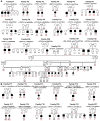
Figure 1
Figure 1
Pedigrees of affected families showing segregation of the biallelic MED27 variants. (A) New families; (B) reported families. Filled symbol = affected. Genotype, where indicated, represents the results of the evaluation of the variants by Sanger sequencing.
Figure 2
Figure 2
In silico modelling of MED27 gene, protein and position of the variants. (A) Chromosome 9 showing position of MED27 on 9q34.13. (B) Schematic diagram of MED27 gene and protein. The location of previously reported variants is indicated by a grey line and new variants by a black line. Introns are not to scale. Exon numbers are according to the canonical transcript (NM_021723.5). Amino acid changes are according to the reference sequence NP_004260.2. (C) Mediator complex (PDB:6W1S) coloured by module, with binding regions marked by dotted ellipses. The MED27 protein is coloured in green and is part of the lower tail module. (D) MED27 with residues that present a pathogenic variant are highlighted. Residues that are hidden are in parentheses. Those in grey are gnomAD variants of interest (for interactive view: https://michelanglo.sgc.ox.ac.UK/r/med27).
Figure 3
Figure 3
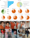
Clinical features of MED27-related disease. (A) The most frequent clinical features. (B) Representative photographs. Top: Patient F3:P1 with generalized dystonia. Bottom from left to right: Patient F3:P2 with flexed and raised arms in elbows with shoulder extension, a suggestion of tongue protrusion; F5:P1 with flexed upper limbs in elbows and wrists and tongue protrusion; F10:P1 with tongue protrusion and cataract, visible in his right eye; F25:P1 with flexion contractures, generalized muscle wasting and a suggestion of jaw-opening dystonia; Patient F24:P4 with generalized dystonia involving the limbs and trunk.
Figure 4
Figure 4
Neuroradiological features of MED27-related disease. Brain MRI images showing the spectrum of basal ganglia anomalies (AE) and cerebellar atrophy (FJ), with a normal subject for comparison (A and F), and of other less frequent neuroimaging findings in the study cohort (KO). (AE) Axial T2-weighted images reveal mild volume reduction of the caudate nuclei in Patient F19:P1 (thin arrows, B); moderate atrophy of both the caudate and putamen nuclei associated with bilateral hyperintensity of the posterior putamen in Patient F24:P7 (arrowheads, C); bilateral moderate atrophy with subtle hyperintensity of the putamen and caudate nuclei in Patient F12:P2 (dashed arrows, D); and severe atrophy with marked hyperintensity of the same nuclei in F3.P1 (thick arrows, E). In addition, Patient F3:P1 shows severe white matter volume loss, with enlargement of the lateral ventricle (asterisks, E) and enlargement of the cerebral subarachnoid spaces. (G and H) Sagittal and coronal T2-weighted images demonstrate mild cerebellar atrophy in Patient F24:P2, with prevalent superior vermian involvement (thin arrow, G) and less severe subarachnoid spaces enlargement of the lateral portions of the cerebellar hemispheres (thick arrows, H). The corpus callosum is slightly thin (open arrow, G). (I and J) Sagittal T1-weighted and coronal T2-weighted images show severe cerebellar atrophy in Patient F20:P2, with prevalent superior vermian involvement (thin arrow, I), marked T2-hyperintensity of the cerebellar cortex (thick arrows, J), pontine hypoplasia (arrowhead, J) and faint T2-hyperintensity of the dentate nuclei (dashed arrows, J). There is moderate periventricular white matter volume loss and T2-hyperintensity (arrowheads, J) with a thin corpus callosum (open arrow, I). (K) Axial T2-weighted image demonstrates volume reduction and faint hyperintensity of the inferior olivary nuclei in Patient F3:P1 (thin arrows). (L) Axial T2-weighted image shows hyperintensity forming a cross in the pons (hot cross bun sign) due to selective degeneration of the transverse pontocerebellar tracts and median pontine raphe nuclei in Patient F20:P2 (thick arrow). Note the presence of bilateral intraocular lens implants (pseudophakia) as a result of the surgical treatment for cataract (arrowheads). (M) Axial diffusion-weighted image reveals bilateral small focal hyperintensities consistent with restricted diffusion at the level of the central midbrain and subthalamic nuclei in Patient F9:P2 (arrowheads). (N) Axial T2-weighted image demonstrates microcephaly with marked simplification of the cortical gyral pattern in Patient F20:P (open arrows). The caudate and putamen nuclei are very small with bilateral focal hyperintensity of the posterior portions of the putamina (arrowheads). (O) Coronal fluid-attenuated inversion recovery image depicts bilateral atrophy and hyperintensity of the olfactory bulbs in Patient F9:P1 (thin arrows).

References

    1. Malik S, Roeder RG. The metazoan mediator co-activator complex as an integrative hub for transcriptional regulation. Nat Rev Genet. 2010;11:761–772. - PMC - PubMed
    1. Allen BL, Taatjes DJ. The mediator complex: A central integrator of transcription. Nat Rev Mol Cell Biol. 2015;16:155–166. - PMC - PubMed
    1. Kaufmann R, Straussberg R, Mandel H, et al. Infantile cerebral and cerebellar atrophy is associated with a mutation in the MED17 subunit of the transcription preinitiation mediator complex. Am J Hum Genet. 2010;87:667–670. - PMC - PubMed
    1. Vodopiutz J, Schmook MT, Konstantopoulou V, et al. MED20 Mutation associated with infantile basal ganglia degeneration and brain atrophy. Eur J Pediatr. 2015;174:113–118. - PubMed
    1. Hashemi-Gorji F, Fardaei M, Tabei SMB, Miryounesi M. Novel mutation in the MED23 gene for intellectual disability: A case report and literature review. Clin Case Rep. 2019;7:331–335.. - PMC - PubMed

Publication types