

Otosclerosis under the magnifying glass
- PMID: 37518876
- PMCID: PMC10520382
- DOI: 10.47162/RJME.64.2.09
Otosclerosis under the magnifying glass
Abstract
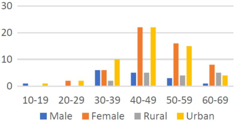
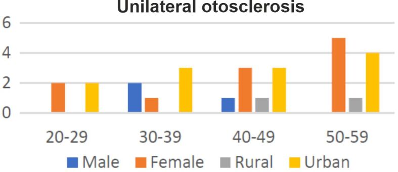
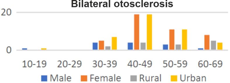
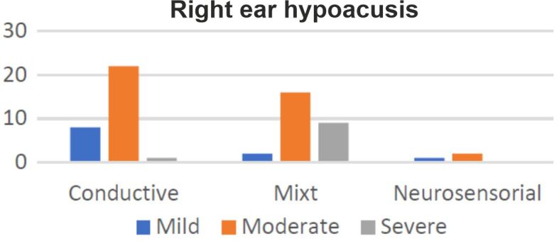
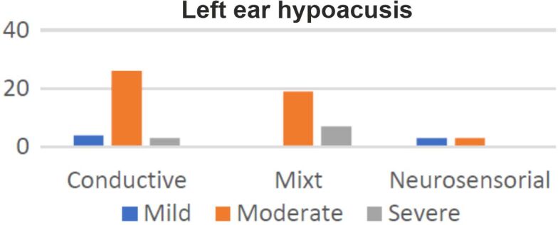
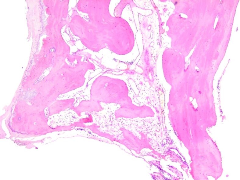
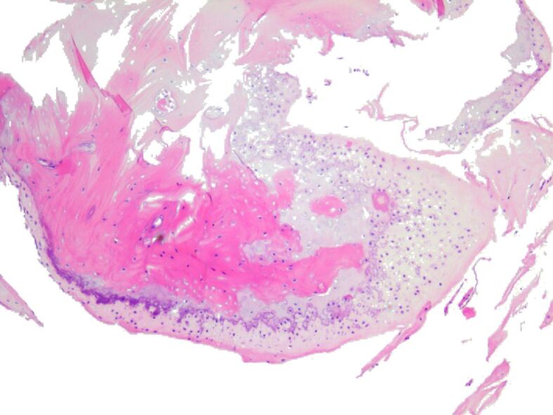
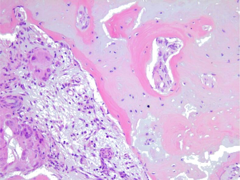
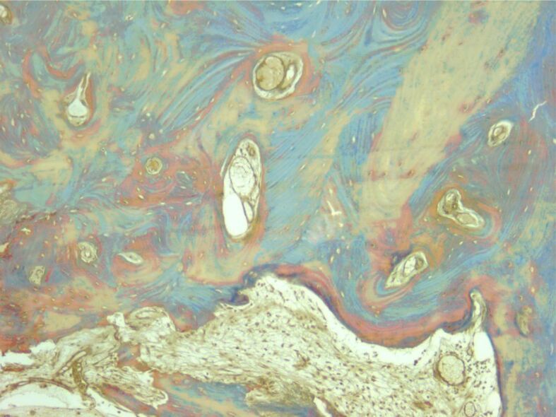
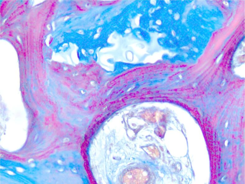
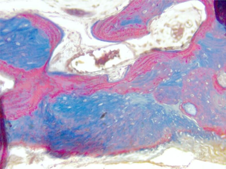
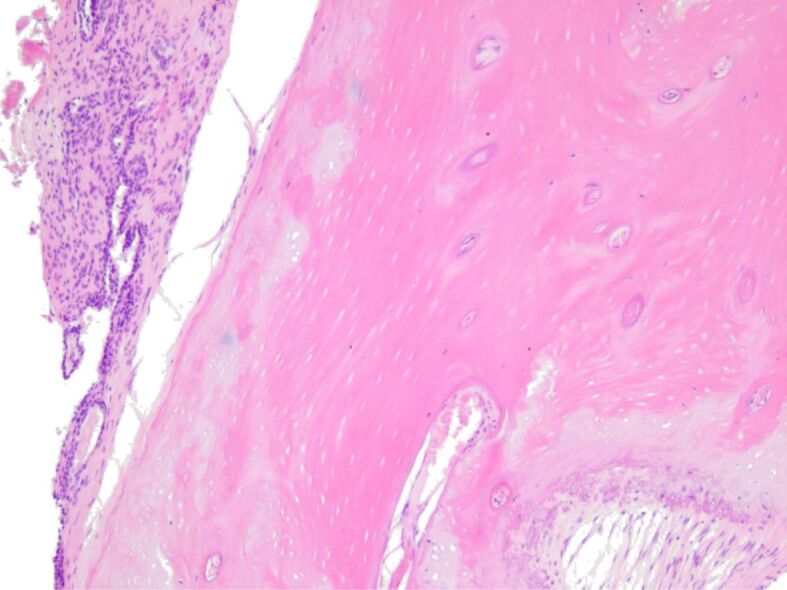
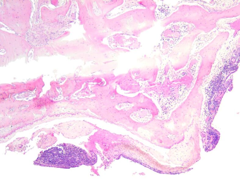
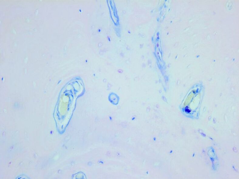
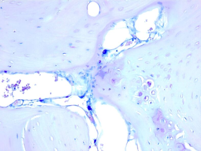
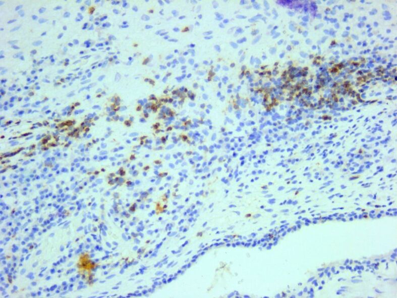
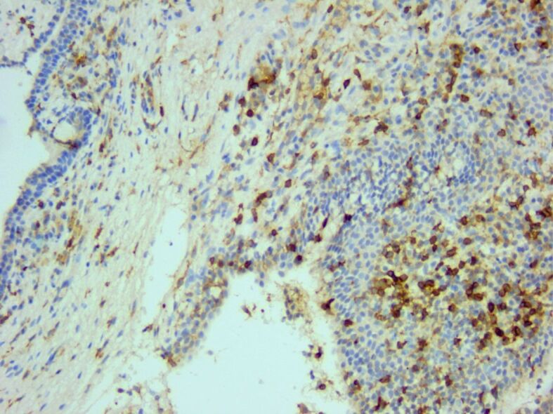
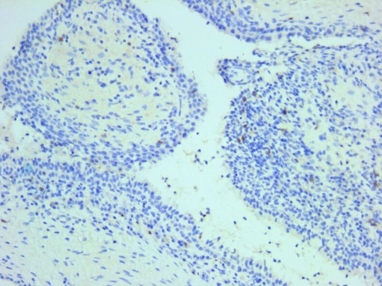
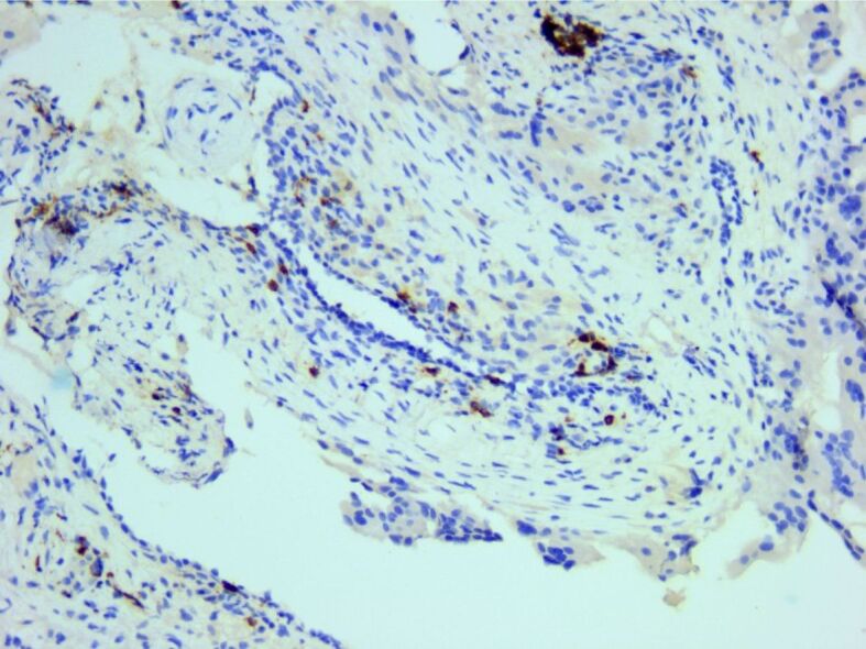
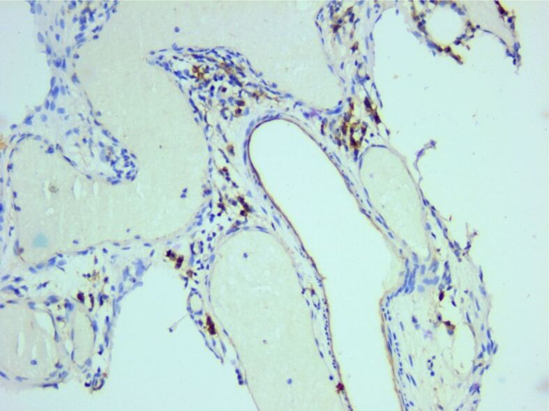
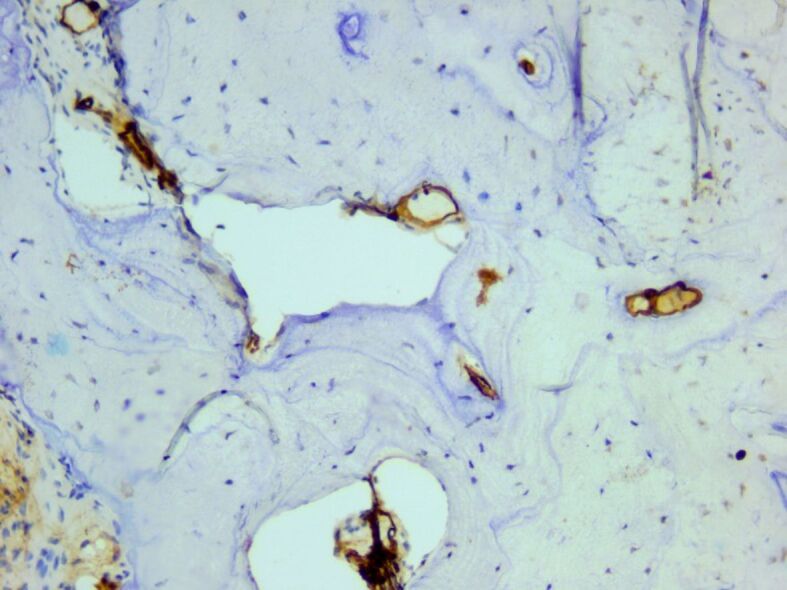
Otosclerosis is a bone condition affecting the stapes bone within the otic capsule, and its exact cause is still unknown. It is characterized by a lack of proper remodeling of newly formed vascular and woven bone, leading to the development of abnormal osteons and the formation of sclerotic bone. Bilateral otosclerosis is seen in 80% of patients and 60% of otosclerosis patients have a family history of the condition. The etiology of this disease is still unknown, there are lots of theories to explain it. The histopathological (HP) studies of otosclerosis showed that osteoblasts, osteoclasts, vascular proliferation, fibroblasts, and histiocytes were observed in the stapes footplate. The onset of the symptoms occurs by the early third decade of life, usually it doesn't start later. In otosclerosis, the energy exerted by sound at the level of the tympanic membrane is reduced in the inner ear due to the fixation and rigidity of the ossicular chain, leading to hearing loss, especially for low frequencies. The primary clinical symptom of otosclerosis is conductive hearing loss but it is important to note that sensorineural hearing loss and mixed hearing loss can also occur as secondary symptoms of the condition. Another symptom present in patients with otosclerosis is tinnitus. The paper carried out a retrospective study of 70 patients diagnosed with otosclerosis in the Department of Otorhinolaryngology of Emergency City Hospital, Timişoara, Romania, between January 2021 to December 2022. Tissue fragments were processed at Service of Pathology by standard Hematoxylin-Eosin staining. The HP diagnosis was completed using Masson's trichrome staining, Giemsa histochemical staining, and immunohistochemical (IHC) reactions with anti-cluster of differentiation (CD)20, anti-CD3, anti-CD4, anti-CD8, anti-CD34, and anti-CD31 antibodies. The microscopic examination showed a chronic diffuse inflammatory infiltrate that consisted predominantly of mature T-lymphocytes, immunohistochemically positive for CD3, CD4 and CD8. There were also present rare CD20-positive B-lymphocytes. Among the lymphocytes, relatively numerous mast cells were identified, highlighted histochemically by the Giemsa staining. They had numerous purple-violet intracytoplasmic granules. In the connective tissue support, a relatively rich vascular network was identified, consisting of hyperemic capillaries, highlighted immunohistochemically with anti-CD31 and anti-CD34 antibodies. Bone tissues trabeculae showed extensive areas of fibrosis. The collagen fibers were highlighted by Masson's trichrome staining, being stained in green, blue, or bluish green.
Conflict of interest statement
The authors declare that they have no conflict of interests.
Figures

References
-
- Rämö JT, Kiiskinen T, Seist R, Krebs K, Kanai M, Karjalainen J, Kurki M, Hämäläinen E, Häppölä P, Havulinna AS, Hautakangas H;, Palta P, Esko T, Metspalu A, Pirinen M, Karczewski KJ, Ripatti S, Milani L, Stankovic KM, Mäkitie A, Daly MJ, Palotie A. Genome-wide screen of otosclerosis in population biobanks: 27 loci and shared associations with skeletal structure. Nat Commun. 2023;14(1):157–157. - PMC - PubMed
-
- Anniko M , Bernal-Sprekelsen M , Bonkowsky V , Bradley PJ , Iurato S , et al., editors. Otorhinolaryngology, head and neck surgery . Berlin-Heidelberg : Springer-Verlag ; 2010 .
-
- Foster MF, Backous DD. Clinical evaluation of the patient with otosclerosis. Otolaryngol Clin North Am. 2018;51(2):319–326. - PubMed
-
- Thys M, Van Camp. Genetics of otosclerosis. Otol Neurotol. 2009;30(8):1021–1032. - PubMed
MeSH terms
LinkOut - more resources
Full Text Sources
Medical
Research Materials
Miscellaneous
