. 2023 Apr-Jun;64(2):199-206.
doi: 10.47162/RJME.64.2.10.

Clinical and morphofunctional identity of the nasal SMAS

Affiliations

Clinical and morphofunctional identity of the nasal SMAS

Marius Valeriu Hînganu et al. Rom J Morphol Embryol. 2023 Apr-Jun.

Abstract

The fascial system of the face (superficial musculo-aponeurotic system, SMAS) in the nasal part is a sustained layer that connects the nearby regions. In this paper, we aimed to emphasize the presence of SMAS in different areas of the nasal region: ala nasi, nasolabial fold, nasal dorsum and radix. We performed three studies (anatomical, histological, and radiological) to demonstrate the existence of nasal SMAS. The study group consisted of cadaveric analyses and retrospective analysis of the patient radiological data. The nasal SMAS was identified as a superficial fascia and a subcutaneous adipose layer. The anatomical dissection study together with histological and radiological evaluations demonstrated the presence of SMAS in the nasal region. We identified peculiarities of the nasal SMAS in two areas: in the ala nasi where it is thinner, and the deep part of the dermis does not adhere to the underlying structures and at the radix and dorsum nasi, where the adipose layer is very thin. The results of our research define nasal SMAS as a unit of great value in facial surgeries, such as facial rejuvenation, the resolution of malformations, or tumor removal.


Conflict of interest statement

The authors declare no conflict of interests.

Figures

Figure 1
Figure 1
Spatial disposition in the anterior regions of the face. 1: The skin layer, epidermis, and dermis, with the denser deep part, serving as the insertion for the muscles of facial expressions; 2: The subcutaneous fat layer; 3: The superficial plane of the facial muscles, which differs according to the studied region; 4: The plane of the branches of the facial nerve – dissection plane medial to the nasolabial groove; 5: The deep plane of the facial muscles, which differs from one region to another – dissection plane lateral to nasolabial groove
Figure 2
Figure 2
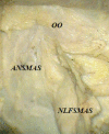
Perioral muscles and nasal region. The levator anguli oris alae nasi (LAON) muscle, SMAS at the ala nasi (ANSMAS), orbicularis oris (OO) muscle, and SMAS in the nasolabial fold (NLFSMAS). Dissection specimen (SOM 62 Kaps microscope, ×20 oculars), 10/1 scale. SMAS: Superficial musculo-aponeurotic system.
Figure 3
Figure 3
Inferior part of the nasolabial groove with the terminal part of the facial artery. Dissection specimen (SOM 62 Kaps microscope, ×20 oculars), 10/1 scale
Figure 4
Figure 4
(a) Skin insertion of the zygomaticus, levator labii superioris, and levator anguli oris muscles – the border area between the zygomatic region (RZ) and cheek (RO); image taken with the operating microscope; (b) Perioral muscles and nasal region; the levator anguli oris alae nasi (LAON) muscle, SMAS at the level of the wing of nose (SN), orbicularis oris (OO) muscle, SMAS at the level of the lower and upper lips (SBIS)
Figure 5
Figure 5
(a) Dissection area lateral and medial to the nasolabial fold; (b) Facial vessels, connective tissue, fat, and fibers of the muscles of the nose. Dissection specimen.
Figure 6
Figure 6
Adhesions and perioral muscles of the nasal region: (a) Ligamentary adhesions that secure the SMAS to the deep fascia; dissection specimen (SOM 62 Kaps microscope, ×20 oculars), 10/1 scale; (b) Conjunctival adipose tissue in the cheek, prebuccinator, and perioral region; dissection specimen
Figure 7
Figure 7
Fibers of the SMAS: (a) SMAS medial to the nasolabial groove, with longitudinal collagen fibers; (b) Thinner collagen fibers and numerous elastic fibers, along with muscle fibers longitudinally arranged in the superior lip of the SMAS; (c) SMAS structure in the angle of the mouth, with numerous interlaced collagen fibers with elastic fibers almost absent; (d) Fibrous attachments that cross the infraSMAS adipose layer in the infraorbital region. Verhoeff’s staining: (a and b) ×400; (c) ×600. Szekely staining: (d) ×400
Figure 8
Figure 8
Various fibers of the SMAS: (a) Elastic fibers of various sizes in the SMAS structure, more numerous on the side of the nasal region; (b) Isolated muscle fibers or small bundles, most of which come from cutaneous muscles that pass through the superficial fascia, medial to the nasolabial groove. Verhoeff’s staining: (a and b) ×400
Figure 9
Figure 9
Graphical representation of quantified percentage volumes in the SMAS in the studied regions.
Figure 10
Figure 10
Axial MRI: (a) Infraorbital insertion and transSMAS of the major zygomatic muscle (ZMI); (b) TransSMAS insertion of the levator labii superioris muscle (TIL). MRI: Magnetic resonance imaging
Figure 11
Figure 11
Specimen sections: (a and c) Sagittal section (SS) through the wing of the nose; SMAS continues to the frontal fascia; (b and d) Axial section (AS) highlighting the jugal SMAS and the insertions into the skin of the nasal muscles, marked with red arrows. A: Anterior insertion of the SMAS on the dorsum nasi.

References

    1. Jost G, Levet Y. Parotid fascia and face lifting: a critical evaluation of the SMAS concept. Plast Reconstr Surg. 1984;74(1):42–51. - PubMed
    1. Mitz V, Peyronie M. The superficial musculo-aponeurotic system (SMAS) in the parotid and cheek area. Plast Reconstr Surg. 1976;58(1):80–88. - PubMed
    1. Hînganu D, Stan CI, Ţăranu T, Hînganu MV. The anatomical and functional characteristics of parotid fascia. Rom J Morphol Embryol. 2017;58(4):1327–1331. - PubMed
    1. Hînganu D, Stan CI, Ciupilan C, Hînganu MV. Anatomical considerations on the masseteric fascia and superficial muscular aponeurotic system. Rom J Morphol Embryol. 2018;59(2):513–516. - PubMed
    1. Hînganu D, Scutariu MM, Hînganu MV. The existence of labial SMAS - anatomical, imaging and histological study. Ann Anat. 2018;218:271–275. - PubMed