. 2023 Apr-Jun;64(2):251-261.
doi: 10.47162/RJME.64.2.16.

Assessment of tumor microenvironment in gastric adenocarcinoma

Affiliations

Assessment of tumor microenvironment in gastric adenocarcinoma

Marius Ionuţ Stancu et al. Rom J Morphol Embryol. 2023 Apr-Jun.

Abstract

Gastric cancer (GC), despite the current possibilities of early diagnosis and curative treatment, remains a major public health problem, being one of the main causes of cancer, due to its detection in advanced stages. Screening programs applied in Western countries led to low incidence rates in these countries. Helicobacter pylori bacterial infection is considered to be the highest risk factor for the onset of GC because it causes chronic inflammation of the gastric mucosa and damages hydrochloric acid secretory glands, eventually leading to atrophic gastritis, which has a potential to progress to GC. In our study, we aimed at assessing the tumor microenvironment in gastric adenocarcinomas as approximately 90% of GCs are adenocarcinomas. Our study showed that the tumor microenvironment has an extremely complex morphological structure, totally different from the microscopic structure of the gastric wall, consisting of stromal cells, lymphocytes, plasma cells, macrophages, blood vessels, collagen fibers, extracellular connective matrix, other cells. The tumor microenvironment presents phenotypic, cellular and molecular heterogeneity; therefore, the microscopic aspect differs from one tumor to another and even from one region to another in the same tumor. Poorly or moderately differentiated adenocarcinomas show a more intense desmoplastic reaction than well-differentiated ones. Alpha-smooth muscle actin (α-SMA)-positive stromal cells (tumor-associated fibroblasts) and tumor macrophages were the most numerous cells of the tumor microenvironment. The tumor microenvironment is the result of cooperation between tumor cells, cancer-associated fibroblasts, immune system cells and blood vessels. It allows tumor cells to multiply, grow and metastasize.


Conflict of interest statement

The authors declare that they have no conflict of interests.

Figures
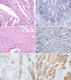
Figure 1
Figure 1
(A) Microscopic image of well-differentiated gastric adenocarcinoma, with multiple cellular and nuclear atypia, and with a low desmoplastic reaction; (B) Gastric adenocarcinoma associated with areas of incomplete intestinal metaplasia and reduced desmoplastic reaction; (C) Well-differentiated gastric adenocarcinoma with intense desmoplastic reaction; (D) Moderately differentiated gastric adenocarcinoma with intense desmoplastic reaction; (E) Well-differentiated gastric adenocarcinoma with low desmoplastic reaction, made up of fine intratumor conjunctive septa with highlighted numerous activated fibroblasts (fibroblasts associated with cancer), intensely positive to the anti-α-SMA antibody; (F) Poorly differentiated gastric adenocarcinoma with intense desmoplastic reaction and numerous activated fibroblasts. HE staining: (A and C) ×200. GS staining: (B and D) ×100. Immunomarking with anti-α-SMA antibody: (E and F) ×200. α-SMA: Alpha-smooth muscle actin; GS: Goldner–Szekely; HE: Hematoxylin–Eosin
Figure 2
Figure 2
Mean values of myofibroblastic cellularity immunomarked with anti-α-SMA antibody, depending on the case. α-SMA: Alpha-smooth muscle actin; C: Case
Figure 3
Figure 3
– (A) Well-differentiated gastric adenocarcinoma associated with a low inflammatory infiltrate, at the level of the tumor stroma; (B) Well-differentiated gastric adenocarcinoma with intense desmoplastic reaction and a moderate inflammatory infiltrate in the tumor stroma; (C) Microscopic image of poorly differentiated gastric adenocarcinoma with intense inflammatory reaction in the tumor stroma; (D) Peritumoral stroma with intense desmoplastic reaction and abundant inflammatory infiltrate. Hematoxylin–Eosin (HE) staining: (A–D) ×200
Figure 4
Figure 4
(A) Heterogeneously distributed T-lymphocytes, mainly in the structure of the conjunctival-vascular septa, but also in the structure of the tumor glands; (B) Peritumoral areas strongly infiltrated with immune cells, predominantly T-lymphocytes. Immunomarking with anti-CD3 antibody: (A and B) ×200. CD3: Cluster of differentiation 3
Figure 5
Figure 5
Mean values of T-lymphocyte cellularity immunomarked with anti-CD3 antibody, depending on the case. C: Case; CD3: Cluster of differentiation 3
Figure 6
Figure 6
(A) Well-differentiated gastric adenocarcinoma with rare B-lymphocytes present in the connective septa between the tumor glands; (B) Moderately differentiated gastric carcinoma with an average number of B-lymphocytes in the tumor stroma. Immunomarking with anti-CD20 antibody: (A and B) ×100. CD20: Cluster of differentiation 20.
Figure 7
Figure 7
Mean values of B-lymphocyte cellularity immunomarked with anti-CD20 antibody, depending on the case. C: Case; CD20: Cluster of differentiation 20
Figure 8
Figure 8
– (A) Poorly differentiated adenocarcinoma infiltrated with numerous macrophages; (B) Numerous macrophages highlighted in the peritumoral stroma. Immunomarking with anti-CD68 antibody: (A and B) ×200. CD68: Cluster of differentiation 68.
Figure 9
Figure 9
Mean values of macrophage cellularity immunomarked with anti-CD68 antibody, depending on the case. C: Case; CD68: Cluster of differentiation 68
Figure 10
Figure 10
(A) Rare plasmocytes identified in the conjunctive septa of the tumor stroma, in a case of well-differentiated gastric adenocarcinoma; (B) Well-differentiated gastric adenoma, with a high number of plasma cells in the tumor stromal septa. Immunomarking with anti-CD79a antibody: (A and B) ×200. CD79a: Cluster of differentiation 79a
Figure 11
Figure 11
Mean values of the plasmocyte cellularity immunomarked with anti-CD79a antibody, depending on the case. C: Case; CD79a: Cluster of differentiation 79a
Figure 12
Figure 12
(A) Well-differentiated gastric adenocarcinoma with a relatively small number of angiogenesis vessels; (B) Moderately differentiated gastric adenocarcinoma with a high number of angiogenesis vessels located mainly around the tumor glands. Immunomarking with anti-CD34 antibody: (A and B) ×200. CD34: Cluster of differentiation 34.
Figure 13
Figure 13
Mean values of the vascular density immunomarked with anti-CD34 antibody, depending on the case. C: Case; CD34: Cluster of differentiation 34

References

    1. Burkitt MD, Duckworth CA, Williams JM, Pritchard DM. Helicobacter pylori-induced gastric pathology: insights from in vivo and ex vivo models. Dis Model Mech. 2017;10(2):89–104. - PMC - PubMed
    1. Sung H, Ferlay J, Siegel RL, Laversanne M, Soerjomataram I, Jemal A, Bray F. Global Cancer Statistics 2020: GLOBOCAN estimates of incidence and mortality worldwide for 36 cancers in 185 countries. CA Cancer J Clin. 2021;71(3):209–249. - PubMed
    1. Forman D, Burley VJ. Gastric cancer: global pattern of the disease and an overview of environmental risk factors. Best Pract Res Clin Gastroenterol. 2006;20(4):633–649. - PubMed
    1. Ferlay J, Shin HR, Bray F, Forman D, Mathers C, Parkin DM. Estimates of worldwide burden of cancer in 2008: GLOBOCAN 2008. Int J Cancer. 2010;127(12):2893–2917. - PubMed
    1. Smyth EC, Nilsson M, Grabsch HI, van Grieken, Lordick F. Gastric cancer. Lancet. 2020;396(10251):635–648. - PubMed