Review
. 2022;31(5):1725-1746.
doi: 10.1007/s11666-022-01402-3. Epub 2022 Apr 22.

Effect of Various Heat Treatments on the Microstructure of 316L Austenitic Stainless Steel Coatings Obtained by Cold Spray

Affiliations
Review

Effect of Various Heat Treatments on the Microstructure of 316L Austenitic Stainless Steel Coatings Obtained by Cold Spray

Laury-Hann Brassart et al. J Therm Spray Technol. 2022.

Abstract

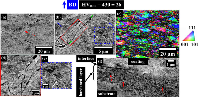
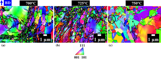
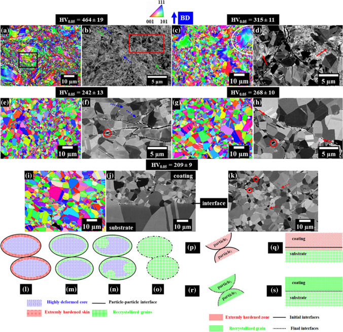
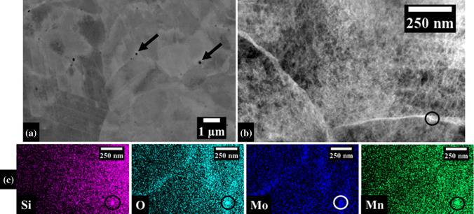
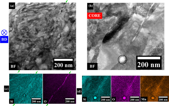
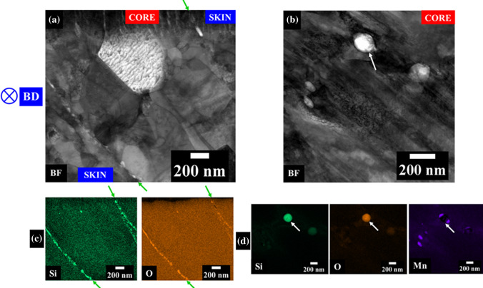
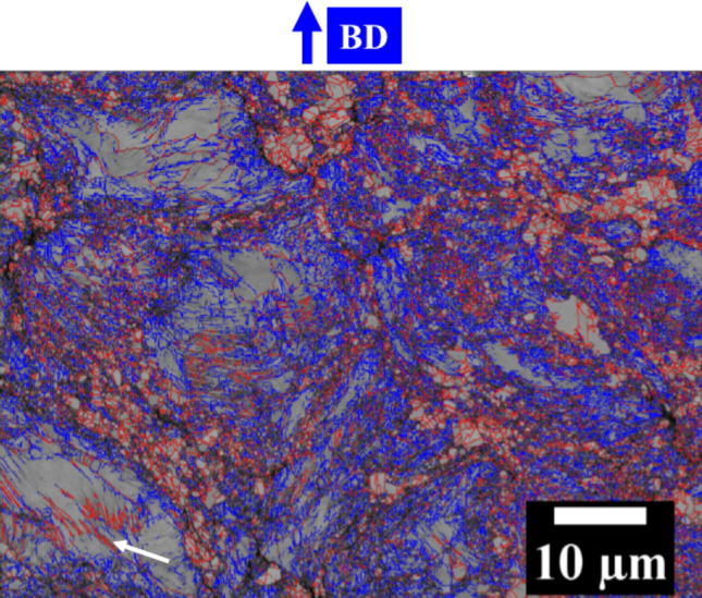
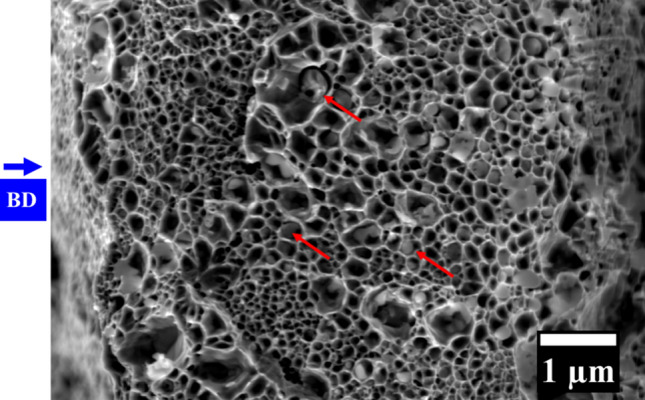
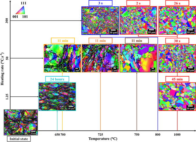
Industries developing cold spray aim at dense and resistant coatings for component repair. However, as-sprayed 316L coatings display non-equilibrium microstructure and brittle fracture behavior. Improving their mechanical properties requires controlling their microstructure; post-spraying heat treatment is a promising approach. The recovery and recrystallization of coatings were little studied, and heat treatments reported in literature mostly used holding for long time in furnaces, not adapted to on-site repairs. This study aimed at gaining insights into recovery and recrystallization mechanisms of 316L coatings, for a broader range of heat treatment kinetics. A study of powders and as-sprayed coatings was conducted to characterize the initial state. In situ XRD measurements provided input for heat treatment definition. Microscopy, room temperature XRD and hardness measurements allowed to better understand the microstructural evolutions and to select treatments leading to original microstructures. In this work, a variety of microstructures were produced by adapting heat treatment conditions for a given set of spraying parameters. The recrystallization path of the heterogeneous skin-core microstructure of deposited particles, as well as the interaction between grain growth and precipitation was revealed. A novel, optimized fast heat treatment led to a fully recrystallized, fine-grained coating and significantly reduced hardness.

Keywords: austenitic stainless steels; cold gas dynamic spraying; heat treatment; microstructure; recrystallization.


Figures

Fig. 1
Fig. 1
(a) Scanning electron micrograph (SEM) of the free feed-stock powder, using Secondary Electron (SE) Everhart–Thornley imaging. (b) Cross-sectional SEM-BSE image of the powder particle microstructure. (c )Cross-sectional SEM-BSE micrograph highlighting: nanosphere precipitates (pointed with arrows), and slip bands (in the red box). (d, e) Respectively, EBSD band contrast (BC) map and inverse pole figure (IPF) map along the y axis of the as-received powder showing the existence of subgrain boundaries (blue arrows) (Color figure online)
Fig. 2
Fig. 2
(a) Curve representing the FWHM evolution of the (111) peak with temperature during in situ XRD. (b) XRD diffractograms of the powder, of the as-sprayed coating and of the D10001min coatings (see Section "Complete recrystallization (F1000, D1000, G100030s)"), with coherence domain sizes (CDS) of each sample indicated close to the corresponding curves
Fig. 3
Fig. 3
(a) Heat cycles for the different heat treatments (logarithmic time scale). (b) Actual heat cycles obtained with the Gleeble system and schematic view of the thermally homogeneous zone (in red) of Gleeble-treated samples. Spikes in the G1000 an G100030s curves are measurement artifacts but did not affect the actually applied heat cycle. BD: Building Direction, C: Coating and S: Substrate (Color figure online)
Fig. 4
Fig. 4
Cross-sectional observations of the as-sprayed (AS) coating. (a) OM image, with some particle boundaries highlighted by blue arrows. (b) SEM-BSE image highlighting some core (dotted square red arrow) and skin (continuous arrow) regions. (c) SEM-BSE micrograph highlighting particle core (dotted square red arrow), skin zone (continuous black arrow), particle boundaries (dashed blue arrow) and nanotwins (dotted green arrow). (d) SEM-BSE micrograph of the substrate–coating interface with an inset showing a magnified zone. e EBSD inverse pole figure map (IPF) along the building direction highlighting the core (bounded by a dotted line circle) and the skin (bounded by a solid line circle) of a particle. (f) TEM bright-field image of the skin zones with particle–particle interface indicated by solid arrows. (g) Detail of (f) with some nano-grains delineated and nanotwins in the square framed region. (h, i) Details of boxes in e indicating nanotwins (in green) in a local image quality map. (j) SEM-BSE image showing the particle–particle interfaces and precipitates in magnified core zones (solid line blue arrows). BD: building direction (Color figure online)
Fig. 5
Fig. 5
Cross-sectional analysis of the F650 sample. (a) SEM-BSE micrograph showing the core-skin structure, highlighting a core region (dotted arrow) and a skin region (continuous arrow). (b) SEM-BSE micrograph highlighting recrystallized grains (framed by continuous lines), nanotwins (framed by dotted lines), PPBs (solid line arrows) and precipitates (dotted line arrows). (c) EBSD IPF map of the coating along the building direction. d Enlarged view of the continuous frame in b, with nano-grains identified by black arrows. (e) Enlarged view of the dotted frame in (b), with nanotwins identified by the white arrow. (f) SEM-BSE micrograph of the substrate–coating interface with some recrystallized grains identified by red arrows (Color figure online)
Fig. 6
Fig. 6
EBSD inverse pole figure maps along BD of (a) D700, (b) D725 and (c) D750 coating samples, with a circled local nano-grained cell
Fig. 7
Fig. 7
(a) EBSD inverse pole figure map along BD of the G800 coating highlighting a core region (bounded by a dotted line circle) and the corresponding skin region (bounded by a solid line circle), with framed nanotwins (thin black lines). (b) Cross-sectional SEM-BSE image of the G800 coating with PPBs indicated by solid lines arrows, framed fine recrystallized grains in the skin zones and precipitates (dotted lines arrows). (c) EBSD inverse pole figure map along BD of the G1000 showing extension of recrystallization and remaining core (circled in dotted line) – skin (circled in solid line). (d) Cross-sectional SEM-BSE image of the G1000 sample showing recrystallization and nanotwins (indicated by arrows). e EBSD inverse pole figure map of the D1000 sample along BD. (f) Cross-sectional SEM-BSE image of the D1000 sample with some PPBs indicated by dotted line arrows and oxide coarsening at PPB triple junctions (circled). (g) EBSD inverse pole figure map along BD of the G100030s heat-treated coating. h Cross-sectional SEM-BSE image of the coating after G100030s treatment showing the recrystallization blocked by PPBs (indicated by solid arrows) and oxide coarsening at PPB triple junctions (circled). (i) EBSD inverse pole figure map along BD of the F1000 sample. (j) Cross-sectional SEM-BSE micrograph of the F1000 substrate–coating interface (pointed by continuous line arrow). (k) Cross-sectional SEM-BSE image of the F1000 coating showing PPB evolution (indicated by arrows) and oxide coarsening at PPB triple junctions (circled). (l, m, n, o) Schematic view of proposed coating recrystallization stages during a post-spraying heat treatment, showing two deposited particles. l as-deposited state; m recrystallization of skin zones; (n) extension of recrystallization into the core region leading to (o) full recrystallization. (p, q, r, s) Schematic view of the evolution stages of particle–particle (p, q) and coating–substrate (r, s) interfaces in a 316L cold sprayed coating during a post-spraying heat treatment up to at least 1000 °C
Fig. 8
Fig. 8
(a) Cross-sectional SEM-BSE micrograph of the as-received powder highlighting nanosphere precipitates (pointed with arrows) (b) TEM high-angle annular dark-field (HAADF) image of an as-received particle with a precipitate circled in solid line. (c) EDS elemental maps of the same zone and silica nanosphere circled in solid lines
Fig. 9
Fig. 9
(a) TEM bright-field image of the as-sprayed coating skin zones with particle–particle interfaces marked by solid arrows. (b) TEM bright-field image of an as-sprayed coating core with a silica nanosphere (in the solid line circle). (c,d) EDS elemental maps on the same zones and silica nanospheres surrounded by a solid line
Fig. 10
Fig. 10
TEM observation of the G800 coating. (a) Bright-field image with particle–particle interfaces indicated by arrows. (b) Bright-field image of a core zone with a silica-rich nanosphere indicated by arrows. (c, d) EDS elemental maps of the zones, respectively, in (a) and (b)
Fig. 11
Fig. 11
Cumulative frequency evolution of the grain size distributions. Values for the substrate grain sizes are to be read on the upper horizontal axis; values for the initial powder and for all coatings are to be read on the lower horizontal axis
Fig. 12
Fig. 12
EBSD image quality map of the G800 heat-treated coating, showing LAGBs (in blue) and HAGBs (in red), with some nanotwins indicated by a solid line arrow
Fig. 13
Fig. 13
SEM secondary electron image of the fracture surface after a tensile test on a fully recrystallized coating (type F1000), highlighting silica-rich precipitates inside fine dimples (red arrows)
Fig. 14
Fig. 14
Summary of microstructural evolution of coatings with applied post-spraying heat treatment. The holding time at maximum temperature is indicated on the top of each EBSD IPF map. Heating rates (to be read along the vertical axis) and holding temperatures (to be read along the horizontal axis) are set point values. Some heat treatment temperatures have been shifted from their location along the horizontal axis, see arrows linking the EBSD maps to the corresponding set point temperatures (one arrow color per value of temperature)

References

    1. H. Assadi, F. Gärtner, T. Stoltenhoff and H. Kreye, Bonding Mechanism in Cold Gas Spraying, Acta Mater., 2003, 51, p 4379-4394. 10.1016/S1359-6454(03)00274-X
    1. S. Adachi and N. Ueda, Effect of Cold-Spray Conditions using a Nitrogen Propellant Gas in AISI 316L Stainless Steel Coating Microstructures, Coat., 2017, 7, p 87-96. 10.3390/coatings7070087
    1. R. Maestracci, Influence de la Microstructure sur les Mécanismes D’endommagement Thermomécanique de Revêtements à base d’acier Inoxydable AISI 316L Réalisés Par Projection Dynamique Par Gaz Froid « cold spray » (Influence of the microstructure on the thermomechanical damage mechanisms of AISI 316L stainless steel coatings produced by cold spray), PhD thesis, MINES ParisTech, 2016, https://pastel.archives-ouvertes.fr/tel-01561419, in French
    1. X. Jiang, N. Overmann, C. Smith and K. Ross, Microstructure, Hardness and Cavitation Erosion Resistance of Different Cold Spray Coatings on Stainless Steel 316 for Hydropower Applications, Mater. Today Commun., 2020, 25, p 101305. 10.1016/j.mtcomm.2020.101305
    1. C. Widener, M. Carter, O. Ozdemir, R. Hrabe, B. Hoiland, T. Stamey, V. Champagne and T.J. Eden, Application of High Pressure Cold Spray for an Internal Bore Repair if a Navy Valve Actuator, J. Therm. Spray Technol., 2016, 25, p 193-201. 10.1007/s11666-015-0366-4

LinkOut - more resources