. 2023 Jun 25;15(6):e40932.
doi: 10.7759/cureus.40932. eCollection 2023 Jun.

The Radiological Spectrum of Rhino-Oculo-Cerebral Mucormycosis

Affiliations

The Radiological Spectrum of Rhino-Oculo-Cerebral Mucormycosis

Dinesh et al. Cureus. .

Abstract

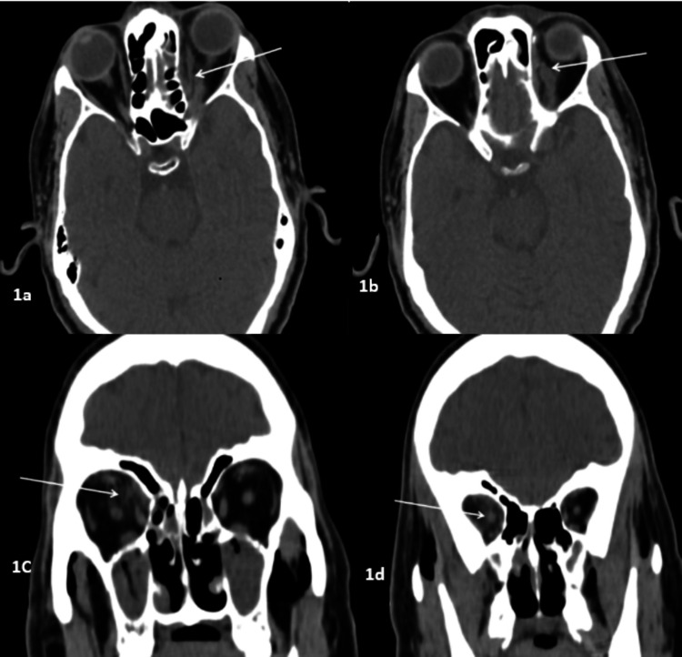
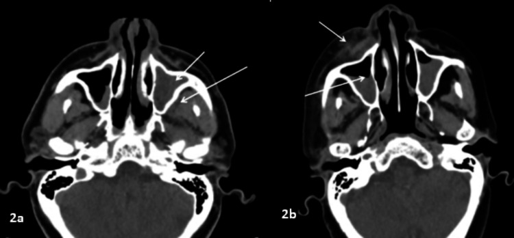
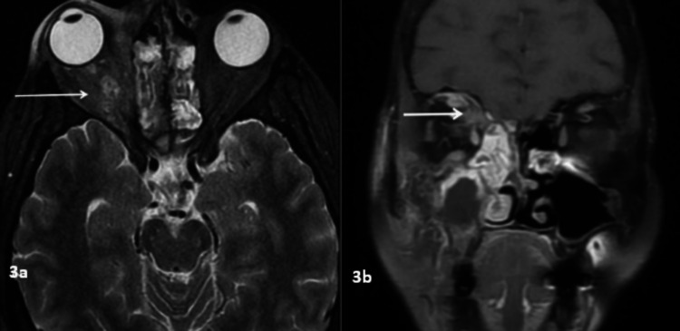
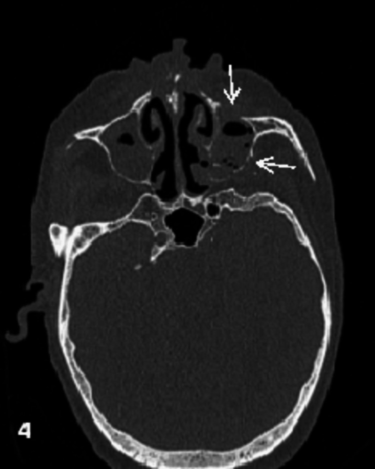
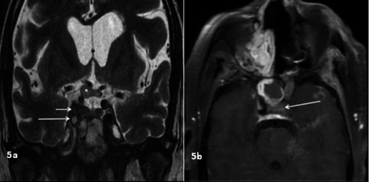
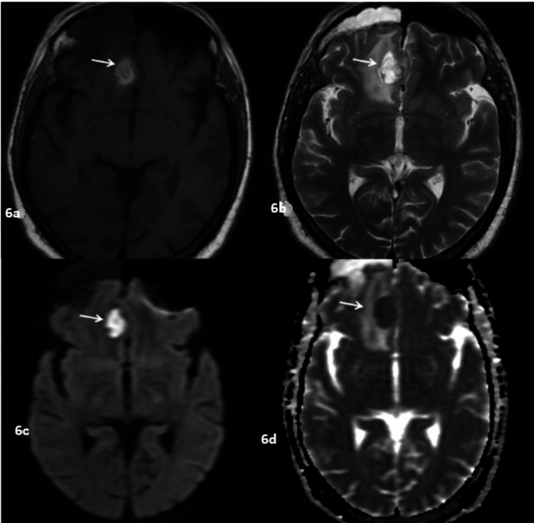
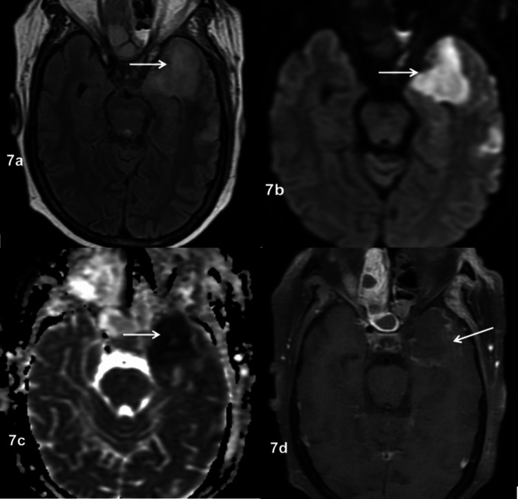
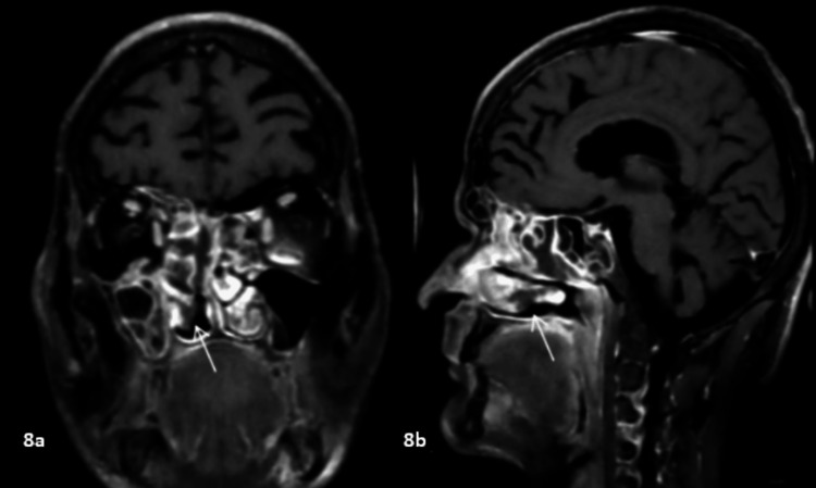
Aim We aim to study the spectrum of imaging findings in patients with rhino-oculo-cerebral mucormycosis (ROCM). Materials and methods This retrospective descriptive study was performed in histopathologically confirmed cases of rhino-oculo-cerebral mucormycosis in a tertiary care center in Bihar, India. The case records of patients with radiological, cultural, and histological evidence of acute invasive ROCM were retrospectively evaluated for relevant radiological and clinical data between May 2021 and June 2022. Results The radiological evaluation included computed tomography (CT) and magnetic resonance imaging (MRI) scans done on 52 patients. The patient's average age was 48 years. The ethmoid sinus was involved in 51 (98%) cases and the maxillary sinus in 50 (96%) cases. Bilateral sinus involvement (45, 86%) was the most common, followed by pansinus involvement (27, 52%). The orbit was involved in 39 (75%) cases, the face in 25 (47%) cases, and retroantral fat stranding in 24 (46%) cases. Mucosal thickening (91%) was the most common pattern of involvement, followed by complete opacification (77%). Osseous involvement was seen in 17 of 44 patients who had CT scans, and the majority of patients had extrasinus extension with intact bone. MRI revealed variable T2SI, with T2 hyperintensity being the most common pattern. Heterogeneous enhancement in post-contrast imaging was the most common. Conclusion ROCM is a life-threatening invasive fungal infection, especially in an immunocompromised state. ROCM is characterized by a variety of imaging abnormalities on CT and MRI, although nonspecific. Imaging aids in suspicion or early diagnosis in appropriate clinical contexts, particularly in an immunocompromised state, and in determining the degree of involvement and complications. Early detection of ROCM and its complications enables proper treatment, which can lower the cost of care, morbidity, and mortality.

Keywords: fungal; mucormycosis infection; paranasal sinus; radiological spectrum; rhino-oculo-cerebral mucormycosis; rocm.


Conflict of interest statement

The authors have declared that no competing interests exist.

Figures

Figure 1
Figure 1. Orbital involvement in different patients
1a and 1b: Axial CT image of a 57-year-old diabetic patient with ROCM showing left intra- and extraconal fat stranding with soft tissue opacification of the left orbital apex (arrow) and thickening of the left superior ophthalmic vein (arrow). 1c and 1d: Coronal CT scan of another 68-year-old patient with ROCM showing right intra- and extraconal fat stranding (arrow) with thickening of the right optic nerve (arrow). CT: computed tomography, ROCM: rhino-oculo-cerebral mucormycosis
Figure 2
Figure 2. Periantral involvement in different patients
2a: Axial CT scan of a 52-year-old patient with mucormycosis showing complete sinus opacification of the left maxillary sinus (short arrow) with retroantral fat stranding (long arrow). 2b: Axial CT scan of a 57-year-old patient with mucormycosis showing air-fluid level in the right maxillary sinus (long arrow) with preantral fat standing (short arrow). Both images show periantral involvement with an intact bony wall, suggestive of the perineural spread of the infection. CT: computed tomography
Figure 3
Figure 3. Orbital involvement
3a: Coronal T2WI of a 43-year-old patient with ROCM showing altered signal intensity in the right intra- and extraconal compartment (arrow). 3b: Coronal post-contrast FS T1WI showing abnormal enhancement of the right intra- and extraconal fat (arrow). T2WI: T2-weighted image, ROCM: rhino-oculo-cerebral mucormycosis, FS T1WI: fat-suppressed T1-weighted image
Figure 4
Figure 4. Osseous involvement
Axial CT bone window image of a 65-year-old diabetic patient who presented with facial pain and redness showing erosion of the anterior and posterior wall of the left maxillary sinus (arrows). CT: computed tomography
Figure 5
Figure 5. Cavernous sinus involvement
5a: Coronal T2WI of a 42-year-old patient who had recent COVID-19 and systemic steroid use who presented with headache and right eye vision loss showing loss of flow of void in the right ICA (large arrow) with abnormal signal intensity in the right cavernous sinus surrounding the right ICA (small arrow) and thickening of the right optic nerve and optic chiasma (asterisk). 5b: Axial post-contrast FS T1WI of a 50-year-old patient who had recent COVID-19 and systemic steroid use who presented with headache and left eye vision loss showing loss of flow void in the left ICA (arrow). T2WI: T2-weighted image, COVID-19: coronavirus disease 2019, ICA: internal carotid artery, FS T1WI: fat-suppressed T1-weighted image
Figure 6
Figure 6. Brain abscess in a 68-year-old diabetic patient with a history of recent systemic steroid use who presented with nasal symptoms and headache
6a: Axial T1WI showing hyperintense lesion in the right gyrus rectus (arrow). 6b: Axial T2WI showing that the lesion is hyperintense on T2WI as well with adjacent edematous changes (arrow). 6c: Axial DWI showing that the lesion shows diffusion restriction (arrow). 6d: ADC map showing that the lesion is hypointense, suggestive of true restriction (arrow). T1WI: T1-weighted image, T2WI: T2-weighted image, DWI: diffusion-weighted imaging, ADC: apparent diffusion coefficient
Figure 7
Figure 7. Subacute infarct in a patient with ROCM
7a: Axial FLAIR image showing hyperintensity in the left frontal lobe (arrow). 7b: Axial DWI at b-1000 showing diffusion restriction in the left frontal lobe (arrow). 7c: ADC map showing hypointensity in the corresponding region (arrow). 7d: Axial post-contrast FS T1WI showing mild peripheral enhancement, suggestive of a subacute infarct (arrow). ROCM: rhino-oculo-cerebral mucormycosis, FLAIR: fluid-attenuated inversion recovery, DWI: diffusion-weighted imaging, ADC: apparent diffusion coefficient, FS T1WI: fat-suppressed T1-weighted image
Figure 8
Figure 8. Black turbinate sign
8a and 8b: Coronal and sagittal post-contrast FS T1WI showing non-enhancing right inferior turbinate (arrows). FS T1WI: fat-suppressed T1-weighted image

Similar articles

Cited by

References

    1. Imaging features of invasive and noninvasive fungal sinusitis: a review. Aribandi M, McCoy VA, Bazan C 3rd. https://doi.org/10.1148/rg.275065189. Radiographics. 2007;27:1283–1296. - PubMed
    1. Imaging features of rhinocerebral mucormycosis: a study of 43 patients. Therakathu J, Prabhu S, Irodi A, Sudhakar SV, Yadav VK, Rupa V. https://doi.org/10.1016/j.ejrnm.2018.01.001 Egypt J Radiol Nucl Med. 2018;49:447–452.
    1. Magnetic resonance imaging in rhino-orbital-cerebral mucormycosis. Sreshta K, Dave TV, Varma DR, Nair AG, Bothra N, Naik MN, Sistla SK. Indian J Ophthalmol. 2021;69:1915–1927. - PMC - PubMed
    1. Code mucor: guidelines for the diagnosis, staging and management of rhino-orbito-cerebral mucormycosis in the setting of COVID-19. Honavar SG. Indian J Ophthalmol. 2021;69:1361–1365. - PMC - PubMed
    1. Epidemiology and clinical manifestations of mucormycosis. Petrikkos G, Skiada A, Lortholary O, Roilides E, Walsh TJ, Kontoyiannis DP. http://dx.doi.org/10.1093/cid/cir866. Clin Infect Dis. 2012;54:0–34. - PubMed

LinkOut - more resources