

The utility of arterial spin labelled perfusion-weighted magnetic resonance imaging in measuring the vascularity of high grade gliomas - A prospective study
- PMID: 37519684
- PMCID: PMC10372548
- DOI: 10.1016/j.heliyon.2023.e17615
The utility of arterial spin labelled perfusion-weighted magnetic resonance imaging in measuring the vascularity of high grade gliomas - A prospective study
Abstract
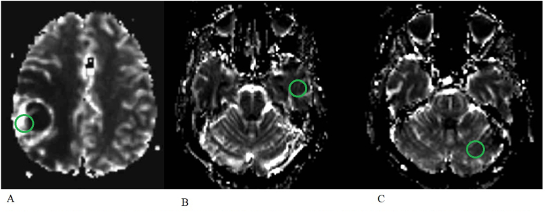
Background: Dynamic susceptibility contrast (DSC) perfusion weighted imaging (PWI) currently remains the gold standard technique for measuring cerebral perfusion in glioma diagnosis and surveillance. Arterial spin labelling (ASL) PWI is a non-invasive alternative that does not require gadolinium contrast administration, although it is yet to be applied in widespread clinical practice. This study aims to assess the utility of measuring signal intensity in ASL PWI in predicting glioma vascularity by measuring maximal tumour signal intensity in patients based on pre-operative imaging and comparing this to maximal vessel density on histopathology.
Methods: Pseudocontinuous ASL (pCASL) and DSC images were acquired pre-operatively in 21 patients with high grade gliomas. The maximal signal intensity within the gliomas over a region of interest of 100 mm2 was measured and also normalised to the contralateral cerebral cortex (nTBF-C), and cerebellum (nTBF-Cb). Maximal vessel density per 1 mm2 was determined on histopathology using CD31 and CD34 immunostaining on all participants.
Results: Using ASL, statistically significant correlation was observed between maximal signal intensity (p < 0.05) and nTBF-C (p < 0.05) to maximal vessel density based on histopathology. Although a positive trend was also observed nTBF-Cb, this did not reach statistical significance. Using DSC, no statistically significant correlation was found between signal intensity, nTBF-C and nTBF-Cb. There was no correlation between maximal signal intensity between ASL and DSC. Average vessel density did not correlate with age, sex, previous treatment, or IDH status.
Conclusions: ASL PWI imaging is a reliable marker of evaluating the vascularity of high grade gliomas and may be used as an adjunct to DSC PWI.
Keywords: Arterial spin labelling (ASL); Cerebral blood flow (CBF); Cerebral blood volume (CBV); Dynamic susceptibility contrast (DSC); Glioma; Magnetic resonance imaging (MRI); Perfusion weighted imaging (PWI).
© 2023 Published by Elsevier Ltd.
Conflict of interest statement
The authors declare that they do not have any competing financial or other interests pertaining to this study.
Figures


References
-
- Louis D.N., Perry A., Wesseling P., Brat D.J., Cree I.A., Figarella-Branger D., Hawkins C., Ng H.K., Pfister S.M., Reifenberger G., Soffietti R., von Deimling A., Ellison D.W. The 2021 WHO classification of tumors of the central nervous system: a summary. Neuro Oncol. 2021 Aug 2;23(8):1231–1251. - PMC - PubMed
-
- Aronen H.J., Gazit I.E., Louis D.N., Buchbinder B.R., Pardo F.S., Weisskoff R.M., Harsh G.R., Cosgrove G.R., Halpern E.F., Hochberg F.H., et al. Cerebral blood volume maps of gliomas: comparison with tumor grade and histologic findings. Radiology. 1994 Apr;191(1):41–51. - PubMed
-
- Chalela J.A., Alsop D.C., Gonzalez-Atavales J.B., et al. Magnetic resonance perfusion imaging in acute ischemic stroke using continuous arterial spin labeling. Stroke. 2000;31:680–687. - PubMed
-
- Kimura H., Kado H., Koshimoto Y., et al. Multislice continuous arterial spin-labeled perfusion MRI in patients with chronic occlusive cerebrovascular disease: a correlative study with CO2 PET validation. J. Magn. Reson. Imag. 2005;22:189–198. - PubMed
LinkOut - more resources
Full Text Sources
Other Literature Sources
