Review
. 2023 Jul 14:10:1172140.
doi: 10.3389/fvets.2023.1172140. eCollection 2023.

Nasal anatomy and sniffing in respiration and olfaction of wild and domestic animals

Affiliations
Review

Nasal anatomy and sniffing in respiration and olfaction of wild and domestic animals

Jinxiang Xi et al. Front Vet Sci. .

Abstract

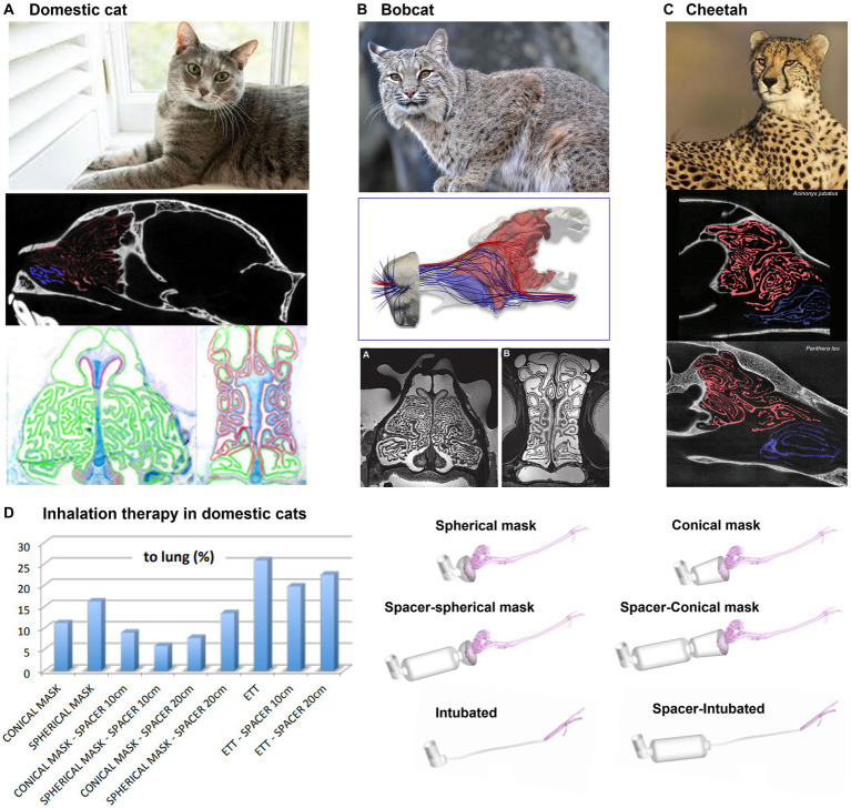
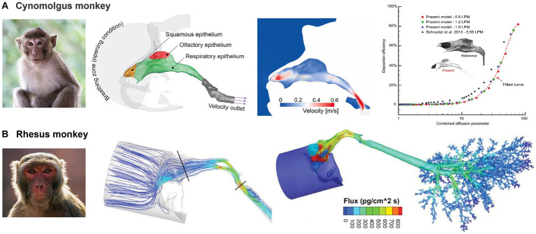
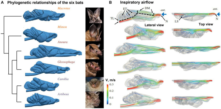
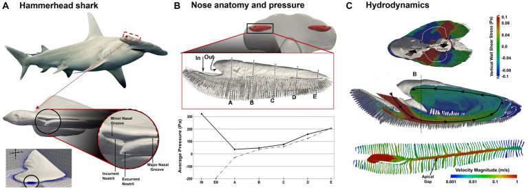
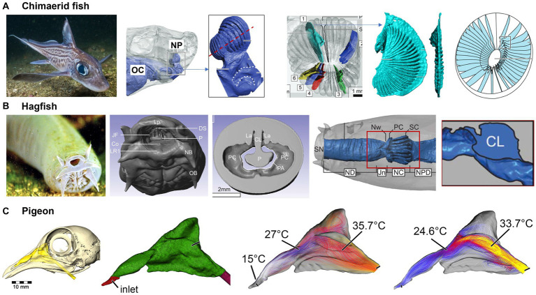
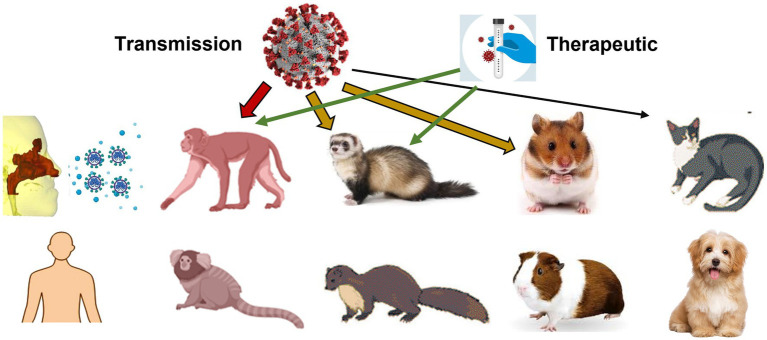
Animals have been widely utilized as surrogate models for humans in exposure testing, infectious disease experiments, and immunology studies. However, respiratory diseases affect both humans and animals. These disorders can spontaneously affect wild and domestic animals, impacting their quality and quantity of life. The origin of such responses can primarily be traced back to the pathogens deposited in the respiratory tract. There is a lack of understanding of the transport and deposition of respirable particulate matter (bio-aerosols or viruses) in either wild or domestic animals. Moreover, local dosimetry is more relevant than the total or regionally averaged doses in assessing exposure risks or therapeutic outcomes. An accurate prediction of the total and local dosimetry is the crucial first step to quantifying the dose-response relationship, which in turn necessitates detailed knowledge of animals' respiratory tract and flow/aerosol dynamics within it. In this review, we examined the nasal anatomy and physiology (i.e., structure-function relationship) of different animals, including the dog, rat, rabbit, deer, rhombus monkey, cat, and other domestic and wild animals. Special attention was paid to the similarities and differences in the vestibular, respiratory, and olfactory regions among different species. The ventilation airflow and behaviors of inhaled aerosols were described as pertinent to the animals' mechanisms for ventilation modulation and olfaction enhancement. In particular, sniffing, a breathing maneuver that animals often practice enhancing olfaction, was examined in detail in different animals. Animal models used in COVID-19 research were discussed. The advances and challenges of using numerical modeling in place of animal studies were discussed. The application of this technique in animals is relevant for bidirectional improvements in animal and human health.

Keywords: COVID-19; animal models; ethmoturbinate; lab animals; livestock; maxilloturbinate; nose function.


Conflict of interest statement

The authors declare that the research was conducted in the absence of any commercial or financial relationships that could be construed as a potential conflict of interest.

Figures

Figure 1
Figure 1
Allometric plots vs. animal body mass: (A) tidal volume (15), (B) respiration frequency (15), and (C) sniff frequency (16). BMR, basal metabolic rate; FMR, field metabolic rate; MMR, maximal metabolic rate.
Figure 2
Figure 2
Canine nasal airway model: (A) MRI images along the axial direction and reconstructed nose model with three regions: vestibule, maxilloturbinate, and ethmoturbinate (39); (B) morphometric parameters (perimeter and cross-sectional area of the sagittal slices), (C) numerical simulations of inspiratory and expiratory flows, (D) sniffing (40), (E) comparison of the pressure patterns between different breeds (Dolicho-, Meso-and Brachycephalic nasal airway and trachea models), and (F) computed resistances at different airway locations (41).
Figure 3
Figure 3
Nasal airway model of a Sprague–Dawley rat (80): (A) life-size and scaled rat nose models of a Sprague–Dawley rat compared to a penny, (B) rat nose and anatomy and different functional regions, (C) numerically predicted airflow field, and (D) precited deposition fraction vs. stokes number (Stk).
Figure 4
Figure 4
Nasal airway model of a New Zealand white rabbit (89): (A) 3D printed and computer model, (B) sagittal MRI images from the nostril to the trachea, (C) reconstructed rabbit nasal airway geometry with different functional regions, and (D) morphometric parameters (perimeter and cross-sectional area of the sagittal slices).
Figure 5
Figure 5
Numerical simulations of rabbit respiration (89, 95): (A) computational mesh with body-fitted prismatic cells and experimental validation, (B) inspiratory airflow streamlines, (C) inspiratory velocity and particle dynamics, and (D) inhalation resistance.
Figure 6
Figure 6
Numerical simulations of rabbit sniffing (95): (A) images of rabbit sniffing, (B) using hyper-mesh to deform the local geometry with prescribed magnitude, (C) deformed front nose with four cross-sectional contours, (D) geometrical effect on nanoparticle deposition in the olfactory region.
Figure 7
Figure 7
Nasal airway anatomy: (A) white-tailed deer (86), (B) sheep (100), and (C) pig (101).
Figure 8
Figure 8
Nasal airway anatomy: (A) racehorse (112), (B) camel with unique thermal/vapor regulation capacity (103).
Figure 9
Figure 9
Felidae nasal airway anatomy: (A) domestic cat, (B) bobcat, (C) cheetah, all with highly intricate ethmoturbinate adapted for acute olfaction (105), and (D) modeling of inhalation therapy in domestic cats (106).
Figure 10
Figure 10
Non-human primate nasal airway models: (A) cynomolgus monkey (107), and (B) rhesus monkey (72).
Figure 11
Figure 11
Phyllostomid bat nasal cavity morphology and olfactory flows (111): (A) phylogenetic relationships of the six bats, and (B) inspiratory flow patterns with lateral and top views.
Figure 12
Figure 12
Hydrodynamics in the nasal region of a hammerhead shark (Sphyrna tudes) (142): (A) head and olfactory chamber, (B) pressure distribution along the incurrent and excurrent channels, and (C) internal flow patterns: surface-limited streamlines and velocity contours.
Figure 13
Figure 13
Fish and bird nasal airways: (A) functional nasal morphology of chimaerid fishes (145), (B) nasal passage of a hagfish (145), and (C) the nasal passage of a pigeon and airflow patterns (146).
Figure 14
Figure 14
Animal models used in COVID research on virus pathogenesis, transmission, and efficacy of treatments and vaccines: primate, ferret, mink, hamster, guinea pig, cat, and dog.

Similar articles

Cited by

References

    1. Mlynski G, Grützenmacher S, Plontke S, Mlynski B, Lang C. Correlation of nasal morphology and respiratory function. Rhinology. (2001) 39:197–201. - PubMed
    1. Raabe OG, Al-Bayati MA, Teague SV, Rasolt A. Regional deposition of inhaled monodisperse coarse and fine aerosol particles in small laboratory animals. Ann Occup Hyg. (1988) 32:53–63. doi: 10.1093/annhyg/32.inhaled_particles_VI.53 - DOI
    1. Gray M, Guido S, Kugadas A. The use of large animal models to improve pre-clinical translational research. Front Vet Sci. (2022) 9:1086912. doi: 10.3389/fvets.2022.1086912 - DOI - PMC - PubMed
    1. Gutting BW, Nichols TL, Channel SR, Gearhart JM, Andrews GA, Berger AE, et al. . Inhalational anthrax (Ames aerosol) in naïve and vaccinated New Zealand rabbits: characterizing the spread of bacteria from lung deposition to bacteremia. Front Cell Infect Microbiol. (2012) 2:87. doi: 10.3389/fcimb.2012.00087, PMID: - DOI - PMC - PubMed
    1. Twenhafel NA. Pathology of inhalational anthrax animal models. Vet Pathol. (2010) 47:819–30. doi: 10.1177/0300985810378112 - DOI - PubMed