. 2023 Sep;47(3):591-607.
doi: 10.1007/s12639-023-01602-w. Epub 2023 Jun 7.

Application of honeybee venom loaded nanoparticles for the treatment of chronic toxoplasmosis: parasitological, histopathological, and immunohistochemical studies

Affiliations

Application of honeybee venom loaded nanoparticles for the treatment of chronic toxoplasmosis: parasitological, histopathological, and immunohistochemical studies

Heba M El Naggar et al. J Parasit Dis. 2023 Sep.

Abstract

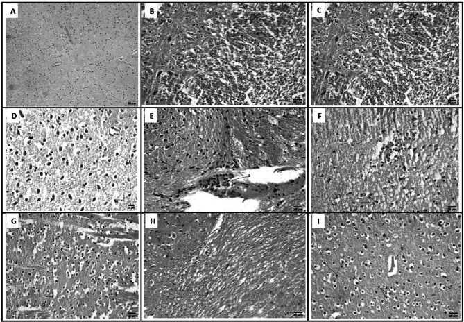
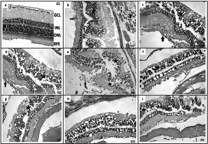
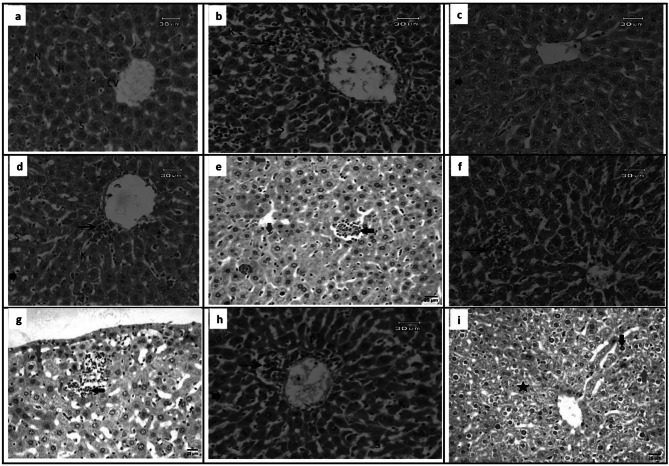
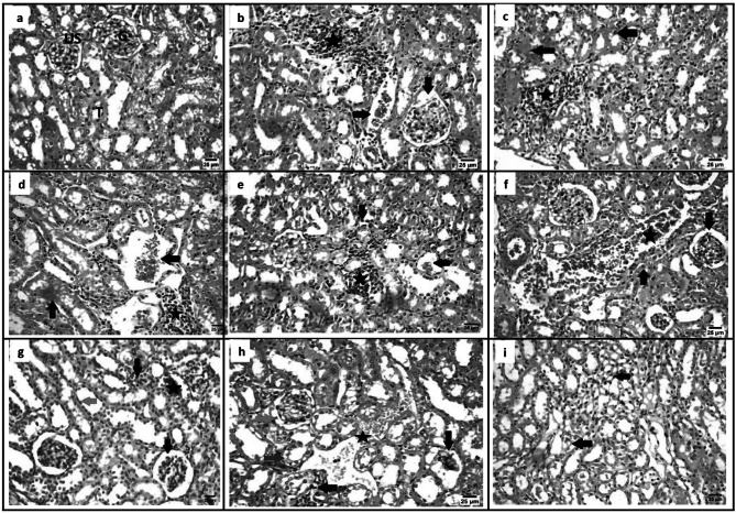
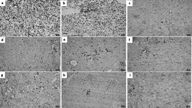
Toxoplasma gondii is an opportunistic intracellular protozoon which may cause severe disease in the immunocompromised patients. Unfortunately, the majority of treatments on the market work against tachyzoites in the acute infection but can't affect tissue cysts in the chronic phase. So, this study aimed to evaluate the effect of bee venom (BV) loaded metal organic frameworks (MOFs) nanoparticles (NPs) for the treatment of chronic murine toxoplasmosis. Ninety laboratory Swiss Albino mice were divided into 9 groups (10 mice each); GI (negative control), GII (infected control), GIII-GXI (infected with Me49 strain of Toxoplasma and treated); GIII (MOFs-NPs), GIV and GV (BV alone and loaded on MOFs-NPs), GVI and GVII (spiramycin alone and loaded on MOFs-NPs), GVIII and GIX (ciprofloxacin alone and loaded on MOFs-NPs). Parasitological examination of brain cyst count, histopathological study of brain, retina, liver, and kidney tissue sections and immunohistochemical (IHC) evaluation of liver was performed. Counting of Toxoplasma brain cysts showed high statistically significant difference between the infected treated groups and GII. GV showed the least count of brain cysts; mean ± SD (281 ± 29.5). Histopathological examination revealed a marked ameliorative effect of BV administration when used alone or loaded MOFs-NPs. It significantly reduced tissue inflammation, degeneration, and fibrosis. IHC examination of liver sections revealed high density CD8+ infiltration in GII, low density CD8+ infiltration in GIII, GVI, GVII, GVIII, and GIX while GIV and GV showed intermediate density CD8+ infiltration. BV is a promising Apitherapy against chronic toxoplasmosis. This effect is markedly enhanced by MOFs-NPs.

Keywords: Bee venom; Ciprofloxacin; Metal organic frameworks; Spiramycin; Toxoplasmosis.


Conflict of interest statement

Conflicts of interestNone.

Figures

Fig. 1
Fig. 1
PXRD patterns of Al-BDC before and after loading
Fig. 2
Fig. 2
Micrographs for the material; a before drug loading, b after drug loading
Fig. 3
Fig. 3
A The equilibrium between the different drug concentrations and the loading amount. B Effect of contact time
Fig. 4
Fig. 4
Brain sections of different study groups (Hematoxylin and Eosin, Scale bar 25 µm, × 400). a GI showing normal histological structure of the brain. b GII showing necrobiotic changes in neurons including vacuolar degeneration in cytoplasm (V) with pyknotic nuclei (P) and presence of axonal degeneration (A). c GIII showing necrobiotic changes in neurons including vacuolar degeneration in cytoplasm (V) with pyknotic nuclei (P) and presence of axonal degeneration (A). d GIV showing improvement of brain tissue with mild vacuolated cytoplasm (V), apoptotic cells (A), pyknotic nuclei (P) observable tissue cyst (star). e GV showing focal gliosis (G) and presence of necrobiotic changes in neurons including vacuolar degeneration in cytoplasm (V) with pyknotic nuclei (P), presence of axonal degeneration (A). f GVI showing focal gliosis (G) and presence of necrobiotic changes in neurons including vacuolar degeneration in cytoplasm (V) with pyknotic nuclei (P), presence of axonal degeneration (A). g GVII showing necrobiotic changes in neurons including vacuolar degeneration in cytoplasm (V) with pyknotic nuclei (P) and presence of axonal degeneration (A). h GVIII showing necrobiotic changes in neurons including vacuolar degeneration in cytoplasm (V) with pyknotic nuclei (P) and presence of axonal degeneration (A). i GIX showing moderate necrobiotic changes in neurons including vacuolar degeneration in cytoplasm (V) with pyknotic nuclei (P) and presence of perivascular edema (E). [GI (normal control), GII (infected control), GIII (infected, NPs treated), GIV (infected, BV treated), GV (infected, BVL-MOFs-NPs), GVI (infected, spiramycin treated), GVII (infected, SL-MOFs-NPs), GVIII (infected, ciprofloxacin treated), GIX (infected, CL-MOFs-NPs)]
Fig. 5
Fig. 5
Eye sections of different study groups (Hematoxylin and Eosin, Scale bar 25 µm, X400). a GI showing normal microscopic picture of retina. b GII showing infiltration of ganglionic cell layer by few number mononuclear inflammatory cells (red arrow), mild disorganization (star) of outer nuclear layer (Photoreceptor cells) and presence of tachyzoites of Toxoplasma (T) in smooth muscle (black arrow). c GIII showing infiltration of vitreous by few number of mononuclear inflammatory cells (black arrow), disorganization (D) of outer nuclear layers (Photoreceptor cells) (star). d GIV showing infiltration of vitreous by few number of mononuclear inflammatory cells (black arrow), disorganization (D) of outer nuclear layers (Photoreceptor cells) (star). e GV showing infiltration of ganglionic cell layer by mononuclear inflammatory cells (arrow), disorganization (stars) of inner and outer nuclear layers (Photoreceptor cells). f GVI showing disorganization (stars) of inner and outer nuclear layers (Photoreceptor cells) (stars). g GVII showing disorganization (star) of outer nuclear layers (Photoreceptor cells). h GVIII showing disorganization (star) of outer nuclear layers (Photoreceptor cells). i GIX showing disorganization (star) of outer nuclear layers (Photoreceptor cells). [GI (normal control), GII (infected control), GIII (infected, NPs treated), GIV (infected, BV treated), GV (infected, BVL-MOFs-NPs), GVI (infected, spiramycin treated), GVII (infected, SL-MOFs-NPs), GVIII (infected, ciprofloxacin treated), GIX (infected, CL-MOFs-NPs)]
Fig. 6
Fig. 6
Liver sections of different study groups (Hematoxylin and Eosin, Scale bar 25–30 µm, X400). a GI showing hepatic architecture. The central vein (CV), hepatocytes (H), blood sinusoids (S) and nucleus (S) were noticed. b GII showing degeneration changes with eosinophilic cytoplasm necrotic areas (arrowhead) associated with focal mononuclear cell infiltration (arrow), and dilated sinusoidal (S), deeply pyknotic nuclei (P)with mild activation of Kupffer cells (K). c GIII showing ameliorative effect with few degeneration (arrowhead), enlargement of sinusoids (S), minimum activation of Kupffer cells (K). d GIV showing ameliorative effect with few inflammatory cells around central vein (arrow), enlargement of sinusoids (S), minimum activation of Kupffer cells (K). e GV showing focal infiltration of mononuclear inflammatory cells between hepatocytes (black arrow) with activation of van kupffer cells (blue arrow) and some hepatocytes showing pyknotic nuclei (red arrow). f GVI showing degeneration, mild necrosis (arrowhead), inflammatory cell infiltration (arrow), enlargement of sinusoids (S), mild activation of Kupffer cells (K) and pyknotic nuclei (P). g GVII showing focal infiltration of mononuclear inflammatory cells between hepatocytes (black arrow) and some hepatocytes showing pyknotic nuclei (red arrow). h GVIII showing degeneration, mild necrosis (arrowhead), inflammatory cell infiltration around central vein (arrow), enlargement of sinusoids (S), mild activation of Kupffer cells (K) and pyknotic nuclei (P). i GIX showing necrobiotic changes in some hepatocytes (star), presence of pyknotic nuclei in others (black arrow) and presence of few numbers of mononuclear inflammatory cells in hepatic sinusoids (red arrow). [GI (normal control), GII (infected control), GIII (infected, NPs treated), GIV (infected, BV treated), GV (infected, BVL-MOFs-NPs), GVI (infected, spiramycin treated), GVII (infected, SL-MOFs-NPs), GVIII (infected, ciprofloxacin treated), GIX (infected, CL-MOFs-NPs)]
Fig. 7
Fig. 7
Kidney sections of different study groups (Hematoxylin and Eosin, Scale bar 25 µm, X400). a GI showing normal structure of kidney including glomerulus (G), urinary space, (US) tubules (T). b GII showing focal infiltration by mononuclear inflammatory cells between renal tubules (star) with congestion of peritubular blood vessels (black arrow) and widening urinary space (red arrow). c GIII showing focal infiltration by mononuclear inflammatory cells between renal tubules (star), some renaltubules showing necrobiotic changes (red arrow) and presence of pyknotic nuclei in renal tubular epithelium (black arrow). d GIV showing focal infiltration by mononuclear inflammatory cells between renal tubules (star) with congestion of peritubular blood vessels (black arrow) and some renaltubules showing necrobiotic changes (red arrow). e GV showing focal infiltration by mononuclear inflammatory cells between renal tubules (star) with hypotrophy in some glomeruli (black arrow) and presence of pyknotic nuclei in renal tubular epithelium (black arrow). f GVI showing degeneration (arrowhead), pyknotic nuclei (P) were observed in tubules epithelial cells. g GVII showing focal infiltration by mononuclear inflammatory cells between renal tubules (black arrow) with congestion of peritubular blood vessels (star), widening urinary space (red arrow) and presence of pyknotic nuclei in renal tubular epithelium (green arrow). h GVIII showing some glomeruli showing hypotrophy (black arrow) and others showing widening urinary space (green arrow), some renal tubules showing degenerative changes (blue arrow), presence of focal infiltration by mononuclear inflammatory cells between renal tubules (red arrow) with congestion of peritubular blood vessels (star). i GIX showing disorganization (D) showing congestion of peritubular blood vessels (black arrow) and presence of pyknotic nuclei in renal tubular epithelium (red arrow). [GI (normal control), GII (infected control), GIII (infected, NPs treated), GIV (infected, BV treated), GV (infected, BVL-MOFs-NPs), GVI (infected, spiramycin treated), GVII (infected, SL-MOFs-NPs), GVIII (infected, ciprofloxacin treated), GIX (infected, CL-MOFs-NPs)]
Fig. 8
Fig. 8
IHC results for liver sections of different study groups (IHC-peroxidase-DAB, X400). a, b GII showing high expression of CD8+. c GIII showing low expression of CD8+. d GIV showing intermediate expression of CD8+. e GV showing intermediate expression of CD8+. f GVI showing low expression of CD8+. g GVII showing low expression of CD8+. h GVIII showing low expression of CD8+. i GIX showing low expression of CD8.+. [GI (normal control), GII (infected control), GIII (infected, NPs treated), GIV (infected, BV treated), GV (infected, BVL-MOFs-NPs), GVI (infected, spiramycin treated), GVII (infected, SL-MOFs-NPs), GVIII (infected, ciprofloxacin treated), GIX (infected, CL-MOFs-NPs)]

References

    1. Abd El-Wahed A, Khalifa S, Sheikh B, et al (2018) Bee venom composition: from chemistry to biological activity. In: Studies in natural products chemistry. Elsevier BV, pp 459–484
    1. Abdelhameed R, Abdel-Gawad H, Elshahat M, Emam H. Cu–BTC@cotton composite: design and removal of ethion insecticide from water. RSC Adv. 2016;6:42324–42333. doi: 10.1039/C6RA04719J. - DOI
    1. Acar S, Keskin-Arslan E, Erol-Coskun H, et al. Pregnancy outcomes following quinolone and fluoroquinolone exposure during pregnancy: a systematic review and meta-analysis. Reprod Toxicol. 2019;85:65–74. doi: 10.1016/J.REPROTOX.2019.02.002. - DOI - PubMed
    1. Adade C, Oliveira I, Pais J, Souto-Padrón T. Melittin peptide kills Trypanosoma cruzi parasites by inducing different cell death pathways. Toxicon. 2013;69:227–239. doi: 10.1016/J.TOXICON.2013.03.011. - DOI - PubMed
    1. Ahmed O, Fahim H, Mahmoud A, Ahmed E. Bee venom and hesperidin effectively mitigate complete Freund’s adjuvant-induced arthritis via immunomodulation and enhancement of antioxidant defense system. Arch Rheumatol. 2017;33:198–212. doi: 10.5606/ARCHRHEUMATOL.2018.6519. - DOI - PMC - PubMed

LinkOut - more resources