

Revolutionizing bone regeneration: advanced biomaterials for healing compromised bone defects
- PMID: 37520216
- PMCID: PMC10376722
- DOI: 10.3389/fragi.2023.1217054
Revolutionizing bone regeneration: advanced biomaterials for healing compromised bone defects
Abstract
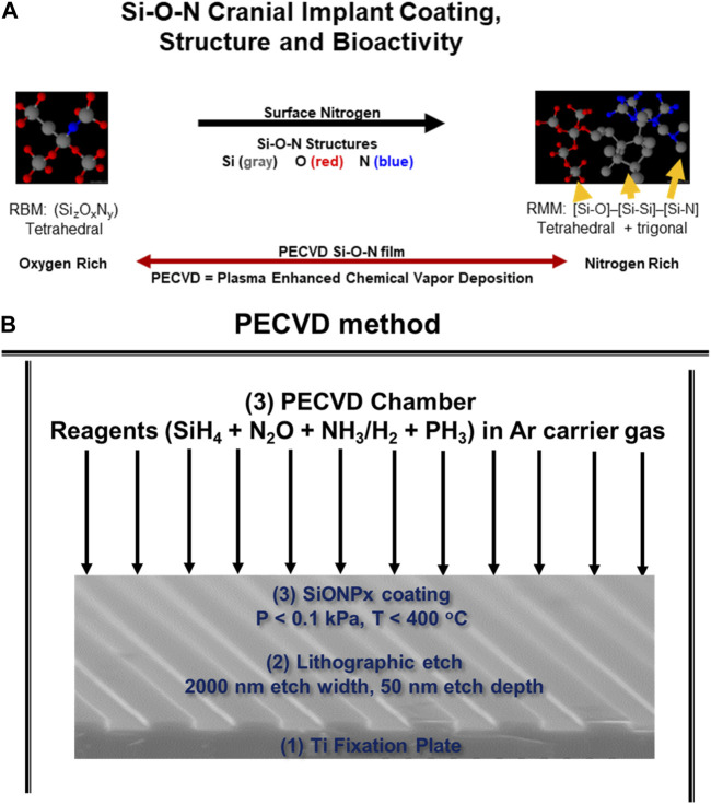
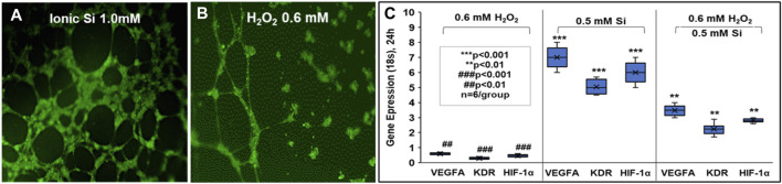
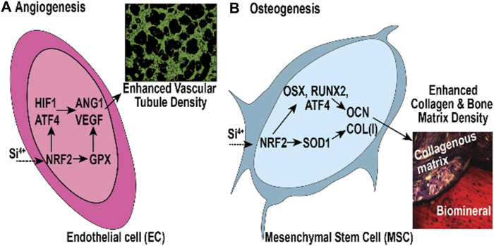
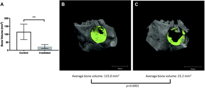
In this review, we explore the application of novel biomaterial-based therapies specifically targeted towards craniofacial bone defects. The repair and regeneration of critical sized bone defects in the craniofacial region requires the use of bioactive materials to stabilize and expedite the healing process. However, the existing clinical approaches face challenges in effectively treating complex craniofacial bone defects, including issues such as oxidative stress, inflammation, and soft tissue loss. Given that a significant portion of individuals affected by traumatic bone defects in the craniofacial area belong to the aging population, there is an urgent need for innovative biomaterials to address the declining rate of new bone formation associated with age-related changes in the skeletal system. This article emphasizes the importance of semiconductor industry-derived materials as a potential solution to combat oxidative stress and address the challenges associated with aging bone. Furthermore, we discuss various material and autologous treatment approaches, as well as in vitro and in vivo models used to investigate new therapeutic strategies in the context of craniofacial bone repair. By focusing on these aspects, we aim to shed light on the potential of advanced biomaterials to overcome the limitations of current treatments and pave the way for more effective and efficient therapeutic interventions for craniofacial bone defects.
Keywords: biomaterials; craniofacial bone defects; engineered biomaterials; oxidative stress; reactive oxygen species; semiconductors.
Copyright © 2023 Awad, Ahuja, Yacoub, Brotto, Young, Mikos, Aswath and Varanasi.
Conflict of interest statement
The authors declare that the research was conducted in the absence of any commercial or financial relationships that could be construed as a potential conflict of interest.
Figures
Similar articles
-
Applications of X-ray computed tomography for the evaluation of biomaterial-mediated bone regeneration in critical-sized defects.J Microsc. 2020 Mar;277(3):179-196. doi: 10.1111/jmi.12844. Epub 2019 Nov 20. J Microsc. 2020. PMID: 31701530 Review.
-
Advances and Prospects in Materials for Craniofacial Bone Reconstruction.ACS Biomater Sci Eng. 2023 Aug 14;9(8):4462-4496. doi: 10.1021/acsbiomaterials.3c00399. Epub 2023 Jul 20. ACS Biomater Sci Eng. 2023. PMID: 37470754 Review.
-
Biomaterials for Craniofacial Bone Regeneration.Dent Clin North Am. 2017 Oct;61(4):835-856. doi: 10.1016/j.cden.2017.06.003. Dent Clin North Am. 2017. PMID: 28886771 Free PMC article. Review.
-
Biomaterials and nanomedicine for bone regeneration: Progress and future prospects.Exploration (Beijing). 2021 Oct 30;1(2):20210011. doi: 10.1002/EXP.20210011. eCollection 2021 Oct. Exploration (Beijing). 2021. PMID: 37323213 Free PMC article.
-
Repair of critical-size porcine craniofacial bone defects using a collagen-polycaprolactone composite biomaterial.Biofabrication. 2021 Nov 1;14(1):10.1088/1758-5090/ac30d5. doi: 10.1088/1758-5090/ac30d5. Biofabrication. 2021. PMID: 34663761 Free PMC article.
Cited by
-
SiONx Coating Regulates Mesenchymal Stem Cell Antioxidant Capacity via Nuclear Erythroid Factor 2 Activity under Toxic Oxidative Stress Conditions.Antioxidants (Basel). 2024 Feb 1;13(2):189. doi: 10.3390/antiox13020189. Antioxidants (Basel). 2024. PMID: 38397787 Free PMC article.
-
Self-assembled peptide hydrogel loaded with functional peptide Dentonin accelerates vascularized bone tissue regeneration in critical-size bone defects.Regen Biomater. 2024 Aug 23;11:rbae106. doi: 10.1093/rb/rbae106. eCollection 2024. Regen Biomater. 2024. PMID: 39263324 Free PMC article.
References
-
- Al-Hezaimi K., Ramalingam S., Al-Askar M., ArRejaie A. S., Nooh N., Jawad F., et al. (2016). Real-time-guided bone regeneration around standardized critical size calvarial defects using bone marrow-derived mesenchymal stem cells and collagen membrane with and without using tricalcium phosphate: An in vivo micro-computed tomographic and histologic experiment in rats. Int. J. Oral Sci. 8, 7–15. 10.1038/ijos.2015.34 - DOI - PMC - PubMed
Publication types
Grants and funding
LinkOut - more resources
Full Text Sources