. 2023 Jul 17:29:214-229.
doi: 10.1016/j.bioactmat.2023.06.007. eCollection 2023 Nov.

Molecular design of an ultra-strong tissue adhesive hydrogel with tunable multifunctionality

Affiliations

Molecular design of an ultra-strong tissue adhesive hydrogel with tunable multifunctionality

Yuting Zheng et al. Bioact Mater. .

Abstract

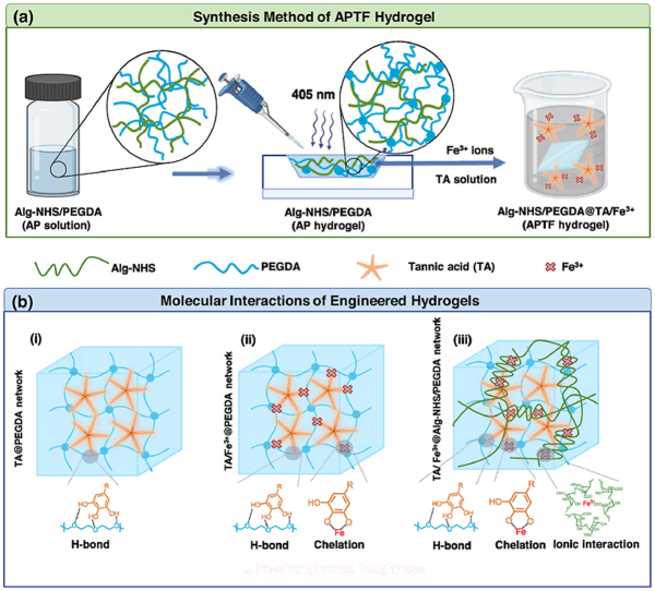
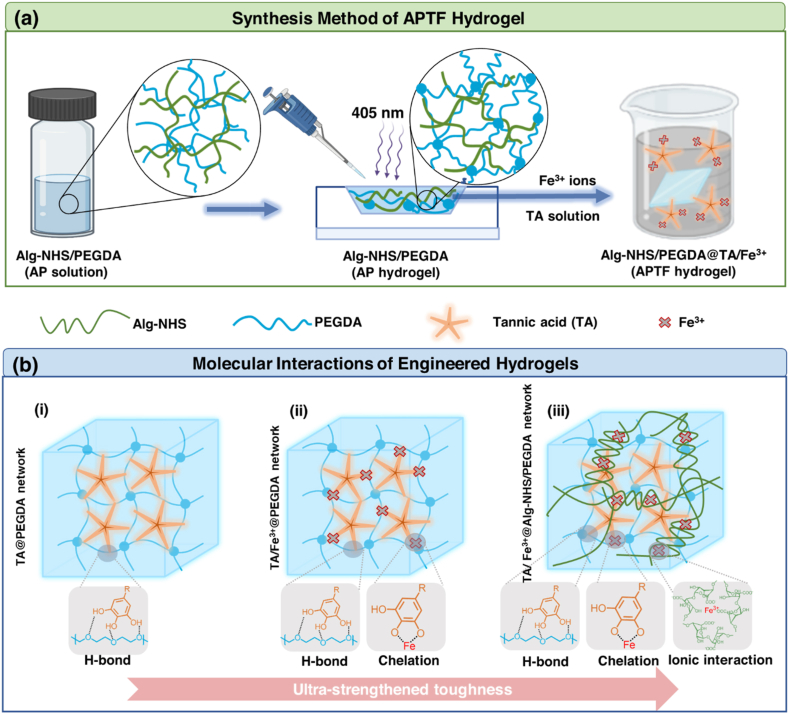
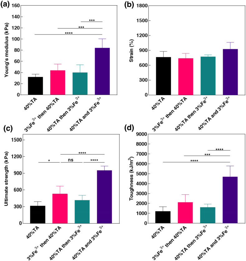
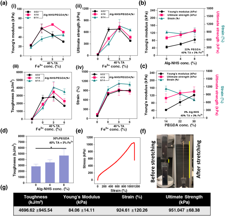
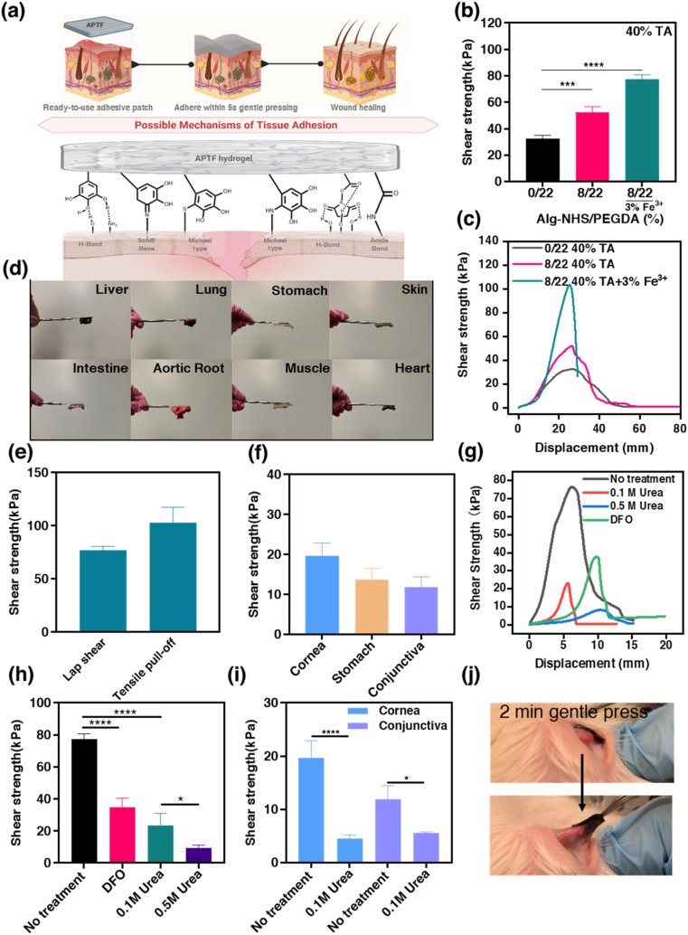
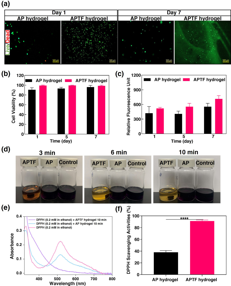
Designing adhesive hydrogels with optimal properties for the treatment of injured tissues is challenging due to the tradeoff between material stiffness and toughness while maintaining adherence to wet tissue surfaces. In most cases, bioadhesives with improved mechanical strength often lack an appropriate elastic compliance, hindering their application for sealing soft, elastic, and dynamic tissues. Here, we present a novel strategy for engineering tissue adhesives in which molecular building blocks are manipulated to allow for precise control and optimization of the various aforementioned properties without any tradeoffs. To introduce tunable mechanical properties and robust tissue adhesion, the hydrogel network presents different modes of covalent and noncovalent interactions using N-hydroxysuccinimide ester (NHS) conjugated alginate (Alg-NHS), poly (ethylene glycol) diacrylate (PEGDA), tannic acid (TA), and Fe3+ ions. Through combining and tuning different molecular interactions and a variety of crosslinking mechanisms, we were able to design an extremely elastic (924%) and tough (4697 kJ/m3) multifunctional hydrogel that could quickly adhere to wet tissue surfaces within 5 s of gentle pressing and deform to support physiological tissue function over time under wet conditions. While Alg-NHS provides covalent bonding with the tissue surfaces, the catechol moieties of TA molecules synergistically adopt a mussel-inspired adhesive mechanism to establish robust adherence to the wet tissue. The strong adhesion of the engineered bioadhesive patch is showcased by its application to rabbit conjunctiva and porcine cornea. Meanwhile, the engineered bioadhesive demonstrated painless detachable characteristics and in vitro biocompatibility. Additionally, due to the molecular interactions between TA and Fe3+, antioxidant and antibacterial properties required to support the wound healing pathways were also highlighted. Overall, by tuning various molecular interactions, we were able to develop a single-hydrogel platform with an "all-in-one" multifunctionality that can address current challenges of engineering hydrogel-based bioadhesives for tissue repair and sealing.

Keywords: Bioadhesive; Molecular engineering; Multifunctionality; Tough hydrogel.


Figures

Image 1
Graphical abstract
Scheme 1
Scheme 1
(a) Schematically illustrated synthesis of APTF hydrogel based on Alg-NHS, PEGDA, and TA/Fe3+. (b) Molecular interactions among the building blocks of APTF hydrogel: (i) TA crosslinked PEGDA network (ii) TA/Fe3+ crosslinked PEGDA network and (iii) TA/Fe3+ crosslinked AP network.
Fig. 1
Fig. 1
Characterizations of synthesized Alg-NHS, AP hydrogel, APTF hydrogel. (a) Schematic for Alg-NHS synthesis. (b)1H NMR and (c) FTIR spectra of Alg, Alg conjugated with NHS under different reaction conditions. FTIR spectra showing (i) carbonyl stretch of NHS (1780 cm−1), (ii) carbonyl stretch of NHS (1704 cm−1), (iii) CNC stretch of NHS (1219 cm−1). (d) The viscosity of synthesized Alg-NHS and Alg at 2%. (e) Rheology characterizations (G′, G″) of Alg/Fe3+, Alg-NHS/Fe3+, and Alg-NHS/TA/Fe3+ hydrogel, the concentrations of Alg, Fe3+ and TA in this study were 2%, 3% and 40% respectively. (f) FTIR spectra of photocrosslinked PEGDA hydrogel (22%) and AP hydrogel consisting of 8% Alg-NHS and 22% PEGDA: (i) carbonyl stretch of NHS (1780 cm−1), (ii) carbonyl stretch of NHS (1704 cm−1), (iii) CNC stretch of NHS (1219 cm−1). (g) XPS survey of PEDGA and APTF hydrogel. (h) Carbon XPS of APTF hydrogel.
Fig. 2
Fig. 2
Physical characterizations of PEGDA, PEGDA/TA, AP, PTF, Alg-NHS/PEGDA hydrogel treated with TA (APT) and APTF hydrogel. (a) Macroscopic images of PEGDA hydrogels crosslinked with TA (40%), and TA/Fe3+ with varied Fe3+ concentrations (3%, 6%). (b) Releasing profile of TA from PEGDA/TA or PTF hydrogels with varied Fe3+ concentrations. (c) UV–Vis of released TA from PEGDA/TA or PTF hydrogels. (d) Representative SEM images of the (i) PEGDA, (ii) AP, (iii) PEGDA/TA, (iv) PTF and (v) APTF hydrogel. Scar bar = 10 μm.
Fig. 3
Fig. 3
Effects of treatment methods on mechanical properties of APTF hydrogel. (a) Young's modulus, (b) strain, (c) ultimate strength, and (d) toughness of APTF hydrogels using different treatment methods for crosslinking.
Fig. 4
Fig. 4
Mechanical characterizations of APTF hydrogels formed by varying Alg-NHS/PEGDA ratios. (a) Mechanical properties of APTF hydrogels with fixed total polymer concentration (22%) and varied treatment methods (40% TA/0%, 3%, 6% Fe3+). (i) Young's modulus, (ii) ultimate strength and (iii) toughness and (iv) ultimate strain of hydrogels treated with TA/Fe3+. Young's modulus, strain, and ultimate strength of APTF hydrogels formed with varied (b) Alg-NHS concentrations and (c) PEGDA concentrations. (d) Toughness of APTF hydrogels with varied Alg-NHS concentrations. (e) Representative stress-strain curve of the optimized engineered APTF hydrogel. (f) Stretching of the APTF hydrogel. (g) Mechanical parameters of optimized APTF hydrogel.
Fig. 5
Fig. 5
Adhesion assessment of APTF hydrogels. (a) Illustration of adhesion mechanism to wet tissue surfaces. (b) Shear strength of different hydrogels on porcine skin and their (c) typical lap shear stress-strain profiles. (d) Demonstration of adhesiveness of APTF patch to different tissues (one side adhered to a spatula and another side adhered to tissue within 5 s gentle pressing). (e) Adhesive strength of APTF hydrogel using two ASTM testing methods including lap shear test and tensile pull-off test. (f) Shear strength of APTF hydrogel on different wet tissues. (g) Representative lap shear stress-strain curve and (h) shear strength of APTF hydrogels on porcine skin with and without urea or DFO treatment. (i) Shear strength of APTF hydrogels on porcine cornea and conjunctiva with and without urea treatment. (j) Demonstration of strong adhesion to rabbit conjunctiva in situ.
Fig. 6
Fig. 6
In vitro biocompatibility and antioxidant activity of APTF hydrogel. In vitro cell studies: (a) Representative live/dead stained images of 3T3 cells on AP hydrogel and APTF hydrogel on day 1 and day 7. (b) Quantification of cellular viability for 3T3 cells seeded on AP and APTF hydrogel over 7 days of culture. (c) Cellular proliferation on AP hydrogel and APTF hydrogel over time based on PrestoBlue assay. In vitro antioxidant activity: (d) The color changes of the three DPPH• solutions containing hydrogel-lacking control, AP hydrogel, APTF hydrogel over time. (e) Absorbance change of DPPH• before and after the reaction. (f) The DPPH• scavenging activities of AP and APTF hydrogels.
Fig. 7
Fig. 7
Photothermal and antibacterial properties of the APTF hydrogels. Temperature enhancement over time for the (a) dry APTF hydrogels and (b) wet APTF hydrogels under irradiation with a NIR laser (808 nm) under different power densities. (c) Photothermal stability of the engineered APTF hydrogel undergoing 10 consecutive heating cycles. (d) Infrared thermal images of the AP and APTF hydrogels under 808 nm irradiation for 6 min. (e) Quantitative antibacterial efficiency against p. aeruginosa under different treatment conditions. (f) Quantitative antibacterial efficiency of APTF hydrogel against p. aeruginosa and MRSA under 808 nm irradiation. (g) Representative images from the p. aeruginosa colonies on the agar plates after treatment with DPBS, DPBS + light, AP hydrogel + light, APTF hydrogel, APTF hydrogel + light. Images were taken from a Zeiss Axio Observer Z1 inverted microscope. Scar bar = 500 μm. (h) Cell viability in the presence of APTF hydrogel after 5 min irradiation.

References

    1. Yuk H., Varela C.E., Nabzdyk C.S., Mao X., Padera R.F., Roche E.T., Zhao X. Dry double-sided tape for adhesion of wet tissues and devices. Nature. 2019;575(7781):169–174. - PubMed
    1. Gao Y., Peng K., Mitragotri S. Covalently crosslinked hydrogels via step-growth reactions: crosslinking chemistries, polymers, and clinical impact. Adv. Mater. 2021;33(25) - PubMed
    1. Jumelle C., Yung A., Sani E.S., Taketani Y., Gantin F., Bourel L., Wang S., Yuksel E., Seneca S., Annabi N., Dana R. Development and characterization of a hydrogel-based adhesive patch for sealing open-globe injuries. Acta Biomater. 2022;137:53–63. - PMC - PubMed
    1. Annabi N., Rana D., Shirzaei Sani E., Portillo-Lara R., Gifford J.L., Fares M.M., Mithieux S.M., Weiss A.S. Engineering a sprayable and elastic hydrogel adhesive with antimicrobial properties for wound healing. Biomaterials. 2017;139:229–243. - PMC - PubMed
    1. Tang J.C., Xi K., Chen H., Wang L.J., Li D.Y., Xu Y., Xin T.W., Wu L., Zhou Y.D., Bian J., Cai Z.W., Yang H.L., Deng L.F., Gu Y., Cui W.G., Chen L. Flexible osteogenic glue as an all-in-one solution to assist fracture fixation and healing. Adv. Funct. Mater. 2021;31(38)