. 2023 Jul 14:14:1223086.
doi: 10.3389/fimmu.2023.1223086. eCollection 2023.

Cytokine response and damages in the lungs of aging Syrian hamsters on a high-fat diet infected with the SARS-CoV-2 virus

Affiliations

Cytokine response and damages in the lungs of aging Syrian hamsters on a high-fat diet infected with the SARS-CoV-2 virus

Gleb Fomin et al. Front Immunol. .

Abstract

Hypertriglyceridemia, obesity, and aging are among the key risk factors for severe COVID-19 with acute respiratory distress syndrome (ARDS). One of the main prognostic biomarkers of ARDS is the level of cytokines IL-6 and TNF-α in the blood. In our study, we modeled hyperglyceridemia and hypercholesterolemia on 18-month-old Syrian hamsters (Mesocricetus auratus). By 18 months, the animals showed such markers of aging as weight stabilization with a tendency to reduce it, polycystic liver disease, decreased motor activity, and foci of alopecia. The high-fat diet caused an increase in triglycerides and cholesterol, as well as fatty changes in the liver. On the third day after infection with the SARS-CoV-2 virus, animals showed a decrease in weight in the groups with a high-fat diet. In the lungs of males on both diets, there was an increase in the concentration of IFN-α, as well as IL-6 in both males and females, regardless of the type of diet. At the same time, the levels of TNF-α and IFN-γ did not change in infected animals. Morphological studies of the lungs of hamsters with SARS-CoV-2 showed the presence of a pathological process characteristic of ARDS. These included bronchointerstitial pneumonia and diffuse alveolar damages. These observations suggest that in aging hamsters, the immune response to pro-inflammatory cytokines may be delayed to a later period. Hypertriglyceridemia, age, and gender affect the severity of COVID-19. These results will help to understand the pathogenesis of COVID-19 associated with age, gender, and disorders of fat metabolism in humans.

Keywords: COVID-19; SARS-CoV-2; Syrian hamster; cytokine response; high-fat diet.


Conflict of interest statement

The authors declare that the research was conducted in the absence of any commercial or financial relationships that could be construed as a potential conflict of interest.

Figures

Figure 1
Figure 1
Body weight change and liver histology of the Syrian hamster on a high-fat diet. (A) Weight dynamics analysis within four months. (B) Liver parenchyma in animals on the RD diet (x400 magnification). (C) In hamsters on the HF diet, foci with microvesicular fatty changes in hepatocytes (yellow arrowheads) were observed (x400 magnification); (D) low-grade inflammatory infiltrate (red arrow) in the area of microvesicular fatty changes (x400 magnification). (E) moderate inflammatory infiltrate (red arrowheads) in the area of macrovesicular changes (yellow arrowheads) (x400 magnification). All histological images with Hematoxylin-Eosin staining.
Figure 2
Figure 2
Diet-induced hypercholesterolemia and hypertriglyceridemia in Syrian hamsters. Biochemical study of blood serum showed an increase in cholesterol (A) and triglycerides (B) in 18-month-old animals. Differences in glucose (C), aspartate aminotransferase (D), and alanine aminotransferase (E) levels were not observed. n = 5 animals per group. P-value was derived using the Mann-Whitney test. Asterisks denote the level of significance observed, as follows: *p ≤ 0.05; **p ≤ 0.01.
Figure 3
Figure 3
Polycystic liver disease in 18-month-old Syrian hamsters. (A) Multi-lobular cysts in the liver (yellow arrowheads). (B) large multi-locular cysts (x40 magnification),. (C) small cysts (x100 magnification). (D) Cyst walls (x400 magnification). All histological images with Hematoxylin-Eosin staining.
Figure 4
Figure 4
Weight dynamics of infected hamsters. Animals on a normal diet (A–C) did not show significant weight loss. Weight was significantly reduced in males (B) and females (D) on a high-fat diet. P-value was derived using one-way ANOVA with Tukey’s multiple comparisons test. Asterisks denote the level of significance observed, as follows: *p ≤ 0.05.
Figure 5
Figure 5
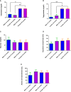
Cytokine response of TNF-α and Il-6 in the lungs of Syrian hamsters on different diets infected with SARS-CoV-2. No changes were found in the level of TNF-α (A, B). And IL-6 levels increased in infected animals (C, D). Cytokine levels were measured in lung homogenates from euthanized animals. n = 5 animals per group. Green bar – healthy animals; Red bar - animals infected with SARS-CoV-2. P-value was derived using one-way ANOVA with Tukey’s multiple comparisons tests. Asterisks denote the level of significance observed, as follows: *p ≤ 0.05; **p ≤ 0.01; ***p ≤ 0.001.
Figure 6
Figure 6
Cytokine response of IFN-α and IFN-γ in the lungs of Syrian hamsters on different diets infected with SARS-CoV-2. Сhanges in interferon levels were noted in males (A–C), but not in females (C, D). Cytokine levels were measured in lung homogenates from euthanized animals. n = 5 animals per group. Green bar – healthy animals; Red bar - animals infected with SARS-CoV-2. P-value was derived using one-way ANOVA with Tukey’s multiple comparisons tests. Asterisks denote the level of significance observed, as follows: *p ≤ 0.05; **p ≤ 0.01; ***p ≤ 0.001.
Figure 7
Figure 7
Histological examination of the liver of Syrian hamsters. (A) low-grade mononuclear infiltrate in the hamster liver parenchyma on the RD diet (x400 magnification). (B) pigment in hamster hepatocytes on the RD diet (blue arrowheads, x400 magnification). (C) - moderately marked granulocyte infiltrate with predominant neutrophils in the liver parenchyma of a hamster on the RD diet infected with SARS-CoV-2 (x400 magnification); (D) Marked multifocal infiltrates (red arrowheads) (x100 magnification) with (E) pigmented macrophages (blue arrowheads) (x400 magnification) in the liver triad region of a hamster infected with SARS-COV-2. n = 6 animals per group. All histological images with Hematoxylin-Eosin staining.
Figure 8
Figure 8
Histological examination of the lungs of Syrian hamsters infected with SARS-CoV-2. (A–C) lungs in healthy animals (x100 magnification -A) (x400 magnification B, C; (D–H) lungs in infected animals: (D) the focus of bronchointerstitial pneumonia (x400 magnification); (E) peribronchial infiltrates and cell debris in the bronchi lumen (x400 magnification); (F) edema and destruction of the pulmonary alveoli (x400 magnification); (G) hemorrhages and destruction of pulmonary alveoli (x400 magnification); (H) intra-alveolar infiltrates and cell debris (x400 magnification). n = 6 animals per group. All histological images with Hematoxylin-Eosin staining.
Figure 9
Figure 9
Scoring of pathological changes in the lungs of SARS-CoV-2 infected animals. There are no significant differences were found between groups of infected animals. A 4-point scale was used: 4 points = extremely severe; 3 points = severe; 2 points = moderate; 1 point = mild; 0 points = no changes. P-value was derived using the Kruskal-Wallis test with Dunn’s multiple comparisons tests.

References

    1. Hu B, Huang S, Yin L. The cytokine storm and COVID-19. J Med Virol (2021) 93:250–6. doi: 10.1002/jmv.26232 - DOI - PMC - PubMed
    1. Mehta P, McAuley DF, Brown M, Sanchez E, Tattersall RS, Manson JJ. COVID-19: consider cytokine storm syndromes and immunosuppression. Lancet (2020) 395:1033–4. doi: 10.1016/S0140-6736(20)30628-0 - DOI - PMC - PubMed
    1. Spadaro S, Park M, Turrini C, Tunstall T, Thwaites R, Mauri T, et al. . Biomarkers for acute respiratory distress syndrome and prospects for personalised medicine. J Inflammation (United Kingdom) (2019) 16:1. doi: 10.1186/s12950-018-0202-y - DOI - PMC - PubMed
    1. Tsai PH, Lai WY, Lin YY, Luo YH, Lin YT, Chen HK, et al. . Clinical manifestation and disease progression in COVID-19 infection. J Chin Med Assoc Wolters Kluwer Health; (2021) 84:3–8. doi: 10.1097/JCMA.0000000000000463 - DOI - PubMed
    1. Dong M, Zhang J, Ma X, Tan J, Chen L, Liu S, et al. . ACE2, TMPRSS2 distribution and extrapulmonary organ injury in patients with COVID-19. Biomed Pharmacother (2020) 131:110678. doi: 10.1016/j.biopha.2020.110678 - DOI - PMC - PubMed

Publication types