. 2023 Jul 10;26(8):107325.
doi: 10.1016/j.isci.2023.107325. eCollection 2023 Aug 18.

circIFNGR2 regulating ankylosing spondylitis-associated inflammation through macrophage polarization

Affiliations

circIFNGR2 regulating ankylosing spondylitis-associated inflammation through macrophage polarization

Minkai Song et al. iScience. .

Abstract

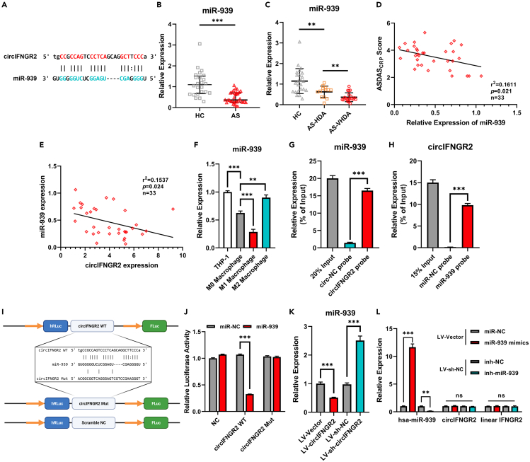
Macrophages activation is crucial in pathogenesis of rheumatic diseases like ankylosing spondylitis (AS). Circular RNAs (circRNAs)-induced macrophage-associated inflammation participates in many autoimmune diseases but remains elusive in AS. Here, we verified increased expression of circIFNGR2 in peripheral blood mononuclear cells from patients with AS and its expression levels were correlated with the AS severity. In vitro assays revealed that circIFNGR2 enhances macrophage proliferation, and regulates M1/M2 macrophage polarization and NF-κB/Akt pathways. We identified that circIFNGR2 promoted the expression of iNOS/TNFα and M1 polarization, and restrained M2 polarization by sponging miR-939. Additionally, the RNA-binding protein, eIF4A3, was found to enhance the production of circIFNGR2. Interestingly, miR-939 attenuated joint damage in collagen-induced arthritis mice, whereas circIFNGR2 reversed this effect. Our findings highlight the pro-inflammatory roles of eIF4A3-induced circIFNGR2 in AS by modulating macrophage-associated inflammation through miR-939.

Keywords: Clinical genetics; Disease; Pathophysiology.


Conflict of interest statement

The authors have declared that no conflict of interest exists.

Figures

None
Graphical abstract
Figure 1
Figure 1
circIFNGR2 was upregulated in PBMCs from patients with AS and correlated with disease activity (A and B) qRT-PCR analysis of the expression of circIFNGR2 in PBMCs from 24 healthy controls and 33 patients with AS including 13 patients with high disease activity (HDA, 2.1≤ASDASCRP score<3.5) and 20 patients with very high disease activity (VHDA, ASDASCRP score≥3.5). (C–E) Correlation analysis of ASDASCRP score, BASDAI score, and serum CRP levels and the expression levels of circIFNGR2 in PBMCs from patients with AS. (F) Schematic illustration of structural characteristics and back-splicing junction of circIFNGR2. (G) The back-splicing junction sequences of circIFNGR2 were validated by Sanger sequencing. (H) RT-PCR amplification of circIFNGR2 and linear IFNGR2 in THP-1 cells treated with or without RNase R. (I) qRT-PCR analysis of the expression level of circIFNGR2, U6, and GAPDH in nuclear-cytoplasm separation assay. Data in A and B are presented as median ± quartile, data in F and K are presented as mean ± SD. ∗∗∗: p<0.001.
Figure 2
Figure 2
circIFNGR2 enhanced M1 macrophage polarization and restrained M2 macrophage polarization (A) qRT-PCR analysis of the expression level of circIFNGR2 in total PBMCs, peripheral CD14+ cells, and peripheral CD14 cells. (B) qRT-PCR analysis of the expression level of circIFNGR2 in THP-1 cells, M0 macrophages, M1 macrophages and M2 macrophages. (C) qRT-PCR analysis of the expression levels of circIFNGR2 and linear IFNGR2 in circIFNGR2 stably transfected THP-1 cells. (D and E) Flow cytometry analysis for EdU proliferation assays in THP-1 cells with overexpression or knockdown of circIFNGR2. (F and G) Flow cytometry analysis of iNOS+TNFα+ cells in THP-1-derived M1 macrophages with overexpression and knockdown of circIFNGR2. (H and I) Western Blot analysis of relative protein expression of iNOS, CD86, IL1β, IL6, and TNFα in THP-1-derived M1 macrophages with overexpression and knockdown of circIFNGR2. (J and K) Western Blot analysis of relative protein expression of cytoplasmic Iκκα/β, total p65 and phosphorylated p65, and nuclear p65 in THP-1-derived M1 macrophages with overexpression and knockdown of circIFNGR2. (L–O) ELISA analysis of the secretion levels of TNFα, IL6, IL1β, and IL23 in THP-1-derived M1 macrophages with overexpression and knockdown of circIFNGR2. (P and Q) Flow cytometry analysis of CD163+CD206+ cells in THP-1-derived M2 macrophages with overexpression and knockdown of circIFNGR2. (R and S) Western Blot analysis of relative protein expression of total mTOR, phosphorylated mTOR, total Akt, and phosphorylated Akt in THP-1-derived M2 macrophages with overexpression and knockdown of circIFNGR2. Data in bar plots are presented as mean ± SD, data in scatterplots are supplemented with line of mean. ∗: p < 0.05; ∗∗: p < 0.01; ∗∗∗: p<0.001; ns: p ≥ 0.05.
Figure 3
Figure 3
eIF4A3 regulated the expression of circIFNGR2 (A) The binding sites of eIF4A3 in the flanking sequences of the IFNGR2 pre-mRNA transcript were predicted by circInteractome. (B) RIP assays for detection of the binding relationships among eIF4A3 and IFNGR2 pre-mRNA transcripts. (C) Western Blot analysis for identification of the binding relationships among eIF4A3 and the flanking sequences of IFNGR2 pre-mRNA transcripts. (D) qRT-PCR analysis of the expression level of eIF4A3 mRNA in THP-1 cells, M0 macrophages, M1 macrophages, and M2 macrophages. (E) qRT-PCR analysis of the expression level of eIF4A3 mRNA, circIFNGR2, and linear IFNGR2 with or without overexpression and knockdown of eIF4A3. (F) qRT-PCR analysis of the expression of circIFNGR2 in PBMCs from 24 healthy controls and 33 patients with AS. (G) Correlation analysis of the expression levels of circIFNGR2 and eIF4A3 mRNA in PBMCs from patients with AS. Data in B, D, and E are presented as mean ± SD, data in F are presented as median and quartiles. ∗∗: p < 0.01; ∗∗∗: p<0.001; ns: p ≥ 0.05.
Figure 4
Figure 4
circIFNGR2 sponged and downregulated the expression of miR-939 (A) Binding sites of circIFNGR2 and miR-939 were predicted by TargetScan. (B and C) qRT-PCR analysis of the expression of miR-939 in PBMCs from 24 healthy controls and 33 patients with AS including 13 patients with high disease activity (HDA, 2.1≤ASDASCRP score<3.5) and 20 patients with very high disease activity (VHDA, ASDASCRP score≥3.5). (D) Correlation analysis of ASDASCRP score and the expression levels of miR-939 in PBMCs from patients with AS. (E) Correlation analysis of the expression levels of miR-939 and circIFNGR2 in PBMCs from patients with AS. (F) qRT-PCR analysis of the expression level of miR-939 in THP-1 cells, M0 macrophages, M1 macrophages, and M2 macrophages. (G) qRT-PCR analysis of miR-939 in the immunoprecipitates of RAP assays using probes for circIFNGR2 or negative control. (H) qRT-PCR analysis of circIFNGR2 in the immunoprecipitates of RAP assays using probes for miR-939 or negative control. (I and J) Dual-luciferase reporter assay for validation of the binding between circIFNGR2 and miR-939. (K) qRT-PCR analysis of the expression level of miR-939 in circIFNGR2 stably transfected THP-1 cells. (L) qRT-PCR analysis of the expression level of miR-939, circIFNGR2, and linear IFNGR2 in THP-1 cells with overexpression or knockdown of miR-939. Data in B and C are presented as median and quartiles, data in bar plots are presented as mean ± SD. ∗∗: p < 0.01; ∗∗∗: p<0.001.
Figure 5
Figure 5
circIFNGR2 regulated the expression of iNOS/TNFα and M1/M2 macrophages polarization via miR-939 (A) Binding sites of circIFNGR2 and 3′ UTR regions of iNOS/TNFα mRNA were predicted by TargetScan. (B and C) qRT-PCR analysis of iNOS/TNFα mRNA in the immunoprecipitates of RAP assays using probes for miR-939 or negative control. (D and E) Western Blot analysis of relative protein expression of iNOS, CD86, IL6, and TNFα in THP-1-derived M1 macrophages with overexpression of circIFNGR2 and miR-939. (F and G) Flow cytometry analysis of iNOS+TNFα+ cells in THP-1-derived M1 macrophages with overexpression of circIFNGR2 and miR-939. (H) ELISA analysis of the secretion levels of TNFα and IL6 in THP-1-derived M1 macrophages with overexpression of circIFNGR2 and miR-939. (I and J) Western Blot analysis of relative protein expression of iNOS, CD86, IL6, and TNFα in THP-1-derived M1 macrophages with knockdown of circIFNGR2 and miR-939. (K and L) Flow cytometry analysis of iNOS+TNFα+ cells in THP-1-derived M1 macrophages with knockdown of circIFNGR2 and miR-939. (M) ELISA analysis of the secretion levels of TNFα and IL6 in THP-1-derived M1 macrophages with knockdown of circIFNGR2 and miR-939. (N–P) Flow cytometry analysis of iNOS+TNFα+ cells in THP-1-derived M2 macrophages with overexpression or knockdown of circIFNGR2 and miR-939. Data in bar plots are presented as mean ± SD, data in scatterplots are supplemented with line of mean. ∗: p < 0.05; ∗∗: p < 0.01; ∗∗∗: p<0.001.
Figure 6
Figure 6
miR-939 ameliorated inflammatory arthritis and restored by circIFNGR2 in vivo (A) Schematic illustration of experimental setup of CIA mouse model induction and transfection of miR-939 and circIFNGR2. (B) Arthritis score of CIA mice with or without overexpression of miR-939 and circIFNGR2. (C) Representative images of hind paws of CIA mice with or without overexpression of miR-939 and circIFNGR2. (D and E) H&E staining and histological score of CIA mice with or without overexpression of miR-939 and circIFNGR2. (F and G) Safranin O-Fast Green (SOFG) staining and Mankin’s score of CIA mice with or without overexpression of miR-939 and circIFNGR2. Data are presented as mean ± SD. ∗: p < 0.05; ∗∗: p < 0.01; ∗∗∗: p<0.001.

Similar articles

References

    1. Taurog J.D., Chhabra A., Colbert R.A. Ankylosing Spondylitis and axial Spondyloarthritis. N. Engl. J. Med. 2016;374:2563–2574. doi: 10.1056/NEJMra1406182. - DOI - PubMed
    1. Mauro D., Thomas R., Guggino G., Lories R., Brown M.A., Ciccia F. Ankylosing spondylitis: an autoimmune or autoinflammatory disease? Nat. Rev. Rheumatol. 2021;17:387–404. doi: 10.1038/s41584-021-00625-y. - DOI - PubMed
    1. Ranganathan V., Gracey E., Brown M.A., Inman R.D., Haroon N. Pathogenesis of ankylosing spondylitis - recent advances and future directions. Nat. Rev. Rheumatol. 2017;13:359–367. doi: 10.1038/nrrheum.2017.56. - DOI - PubMed
    1. Ma W.T., Gao F., Gu K., Chen D.K. The role of monocytes and macrophages in autoimmune diseases: a comprehensive review. Front. Immunol. 2019;10:1140. doi: 10.3389/fimmu.2019.01140. - DOI - PMC - PubMed
    1. Funes S.C., Rios M., Escobar-Vera J., Kalergis A.M. Implications of macrophage polarization in autoimmunity. Immunology. 2018;154:186–195. doi: 10.1111/imm.12910. - DOI - PMC - PubMed