. 2023 Jul 8;26(8):107352.
doi: 10.1016/j.isci.2023.107352. eCollection 2023 Aug 18.

Structural and energetic insights into Mn-to-Fe substitution in the oxygen-evolving complex

Affiliations

Structural and energetic insights into Mn-to-Fe substitution in the oxygen-evolving complex

Masahiro Saito et al. iScience. .

Abstract

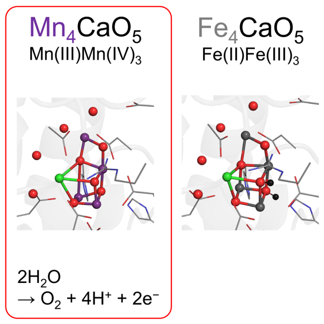
Manganese (Mn) serves as the catalytic center for water splitting in photosystem II (PSII), despite the abundance of iron (Fe) on earth. As a first step toward why Mn and not Fe is employed by Nature in the water oxidation catalyst, we investigated the Fe4CaO5 cluster in the PSII protein environment using a quantum mechanical/molecular mechanical (QM/MM) approach, assuming an equivalence between Mn(III/IV) and Fe(II/III). Substituting Mn with Fe resulted in the protonation of μ-oxo bridges at sites O2 and O3 by Arg357 and D1-His337, respectively. While the Mn4CaO5 cluster exhibits distinct open- and closed-cubane S2 conformations, the Fe4CaO5 cluster lacks this variability due to an equal spin distribution over sites Fe1 and Fe4. The absence of a low-barrier H-bond between a ligand water molecule (W1) and D1-Asp61 in the Fe4CaO5 cluster may underlie its incapability for ligand water deprotonation, highlighting the relevance of Mn in natural water splitting.

Keywords: Catalysis; Chemistry; Photoabsorption.


Conflict of interest statement

The authors declare no competing interest.

Figures

None
Graphical abstract
Figure 1
Figure 1
Protonation state of CP43-Arg357 and D1-His337 and overview of the clusters (A) Fe4CaO5 cluster in Fe(II)Fe(III)3. (B) Mn4CaO5 cluster in Mn(III)Mn(IV)4 (open-cubane S2 conformation). (C) Overview of the Fe4CaO5 cluster. (D) Overview of the Mn4CaO5 cluster. Only H atoms of CP43-Arg357 and D1-His337 are explicitly shown (black ball) for clarity. The black thick arrow indicates the protonation event. Red arrows indicate movement of protons during QM/MM calculations.
Figure 2
Figure 2
Potential energy profile for the O5 position along the Fe1 … O5 … Fe4 and Mn1 … O5 … Mn4 axes (A) Fe4CaO5 cluster. (B) Mn4CaO5 cluster. In the Mn4CaO5 cluster, the global energy minimum corresponds to the open-cubane S2 conformation, whereas the local energy minimum corresponds to the closed-cubane S2 conformation.
Figure 3
Figure 3
Potential energy profile for the H-bond between H2O at W1 and D1-Asp61 (A) Fe4CaO5 cluster. (B) Mn4CaO5 cluster. Labels W1 and Asp61 indicate the W1 and D1-Asp61 moieties in the H-bond, respectively.
Figure 4
Figure 4
H-bond network of the Fe4CaO5 and Mn4CaO5 clusters (A) H-bond network of TyrZ for the Fe4CaO5 cluster. (B) H-bond network of TyrZ for the Mn4CaO5 cluster (open-cubane S2 conformation). (C) H-bond network near the Cl-1 binding site for the Fe4CaO5 cluster. (D) H-bond network near the Cl-1 binding site for the Mn4CaO5 cluster (open-cubane S2 conformation). Dotted lines indicate H-bonds.
Figure 5
Figure 5
Potential energy profile for the H-bond between TyrZ and D1-His190 (A) Fe4CaO5 cluster. (B) Mn4CaO5 cluster. Labels TyrZ and His190 indicate the TyrZ and D1-His190 moieties in the H-bond, respectively. Although the shape of the potential-energy curve for the TyrZ … His190 of the Mn4CaO5 cluster (B) resembles that for the W1 … D1-Asp61 of the Mn4CaO5 cluster (Figure 3B) due to low-barrier H-bonds, the numerical data are different.

Similar articles

Cited by

References

    1. Shen J.R. The structure of photosystem II and the mechanism of water oxidation in photosynthesis. Annu. Rev. Plant Biol. 2015;66:23–48. doi: 10.1146/annurev-arplant-050312-120129. - DOI - PubMed
    1. Umena Y., Kawakami K., Shen J.-R., Kamiya N. Crystal structure of oxygen-evolving photosystem II at a resolution of 1.9 Å. Nature. 2011;473:55–60. - PubMed
    1. Gatt P., Petrie S., Stranger R., Pace R.J. Rationalizing the 1.9 Å crystal structure of photosystem II--a remarkable Jahn-Teller balancing act induced by a single proton transfer. Angew. Chem. Int. Ed. Engl. 2012;51:12025–12028. - PubMed
    1. Kok B., Forbush B., McGloin M. Cooperation of charges in photosynthetic O2 evolution-I. A linear four step mechanism. Photochem. Photobiol. 1970;11:457–475. - PubMed
    1. Saito K., Rutherford A.W., Ishikita H. Energetics of proton release on the first oxidation step in the water-oxidizing enzyme. Nat. Commun. 2015;6:8488. doi: 10.1038/ncomms9488. - DOI - PMC - PubMed