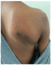
Osteochondroma of Dorsal Scapula: A Case Report and Review of Literature
- PMID: 37521381
- PMCID: PMC10379260
- DOI: 10.13107/jocr.2023.v13.i07.3772
Osteochondroma of Dorsal Scapula: A Case Report and Review of Literature
Abstract
Introduction: Osteochondroma of the scapula constitutes only 3-5% of all osteochondromas; osteochondroma on dorsal aspect of scapula is a rare entity. Diagnosis is almost always clinicoradiologically. Additional computed tomography scan and magnetic resonance imaging may be required for osteochondroma of flat bones such as scapula. Indications for surgery include pain, deformity, dysfunction, neural or vascular compromise, failure of conservative management, or in clinical settings with the high suspicion of malignant transformation and occasionally cosmesis. Outcome of a surgery should be assessed by Patient-Reported Outcome Measures (PROMs) which appraises what "matters to the patient."
Case report: A 10-year-old boy presented to us with painless swelling over the right upper back since 3 years of age and discomfort over the area while sleeping on his back for 6 months. Diagnosis confirmed it to be a pedunculated osteochondroma arising from the dorsal scapula. Here, we report the diagnosis, treatment, and successful Patient-Reported Outcome using QuickDASH© score for an osteochondroma of dorsal scapula using CARE© case reporting guidelines.
Conclusion: We report a rare site of osteochondroma, review the relevant literature, and also stress upon the necessity of analyzing PROMs after surgical treatment of benign tumors of bone which would enable us to evaluate the result of surgery on symptoms, functioning, and health-related quality of life from the patient's perspective.
Keywords: Osteochondroma; Patient Reported Outcome Measure; Patient-Reported Outcome; osteocartilaginous exostosis; scapula.
Copyright: © Indian Orthopaedic Research Group.
Conflict of interest statement
Conflict of Interest: Nil
Figures


References
-
- Cooley LH, Torg JS. “Pseudowinging”of the scapula secondary to subscapular osteochondroma. Clin Orthop Relat Res. 1982;162:119–24. - PubMed
-
- Tomo H, Ito Y, Aono M, Takaoka K. Chest wall deformity associated with osteochondroma of the scapula:A case report and review of the literature. J Shoulder Elbow Surg. 2005;14:103–6. - PubMed
-
- Frost NL, Parada SA, Manoso MW, Arrington E, Benfanti P. Scapular osteochondromas treated with surgical excision. Orthopedics. 2010;33:804. - PubMed
-
- Calvert M, Kyte D, Price G, Valderas JM, Hjollund NH. Maximising the impact of patient reported outcome assessment for patients and society. BMJ. 2019;364:k5267. - PubMed
Publication types
LinkOut - more resources
Full Text Sources
