. 2023 Jul 20:22:100736.
doi: 10.1016/j.mtbio.2023.100736. eCollection 2023 Oct.

Oriented artificial niche provides physical-biochemical stimulations for rapid nerve regeneration

Affiliations

Oriented artificial niche provides physical-biochemical stimulations for rapid nerve regeneration

Minhong Tan et al. Mater Today Bio. .

Abstract

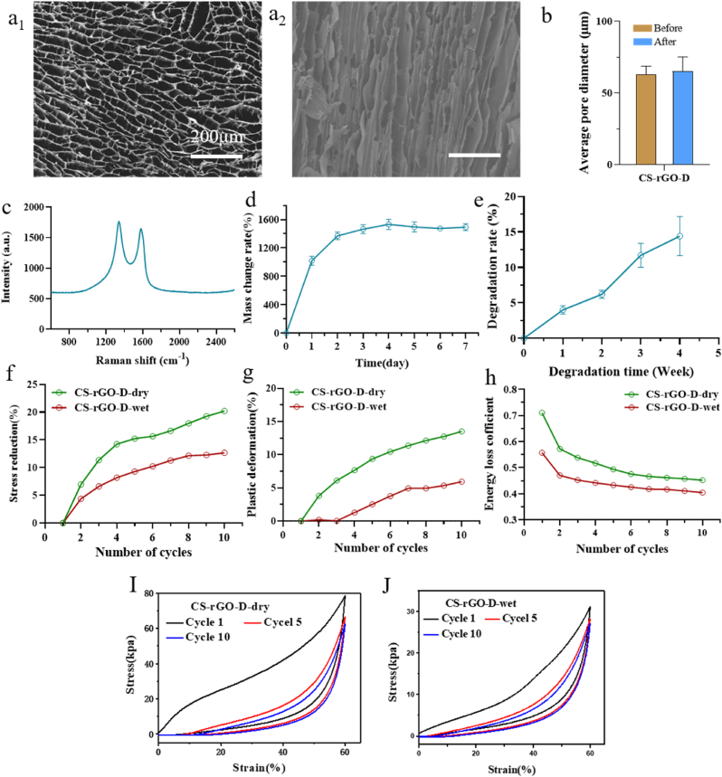
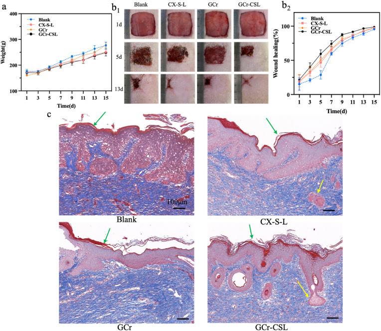
Skin wound is always accompanied with nerve damage, leading to significant sensory function loss. Currently, the functional matrix material based stem cell transplantation and in situ nerve regeneration are thought to be effective strategies, of which, how to recruit stem cells, retard senescence, and promote neural differentiation has been obstacle to be overcome. However, the therapeutic efficiency of the reported systems has yet to be improved and side effect reduced. Herein, a conduit matrix with three-dimensional ordered porous structures, regular porosity, appropriate mechanical strength, and conductive features was prepared by orienting the freezing technique, which was further filled with neural-directing exosomes to form a neural-stimulating matrix for providing hybrid physical-biochemical stimulations. This neural-stimulating matrix was then compacted with methacrylate gelatin (GelMA) hydrogel thin coat that loaded with chemokines and anti-senescence drugs, forming a multi-functional artificial niche (termed as GCr-CSL) that promotes MSCs recruitment, anti-senescence, and neural differentiation. GCr-CSL was shown to rapidly enhances in situ nerve regeneration in skin wound therapy, and with great potential in promoting sensory function recovery. This study demonstrates proof-of-concept in building a biomimetic niche to organize endogenous MSCs recruitment, differentiation, and functionalization for fast neurological and sensory recovery.

Keywords: In situ nerve regeneration; Mesenchymal stem cells; Neural differentiation; Oriented artificial niche; Physical-biochemical factors.


Conflict of interest statement

The authors declare that they have no known competing financial interests or personal relationships that could have appeared to influence the work reported in this paper.

Figures

Image 1
Graphical abstract
Fig. 1
Fig. 1
Scheme of the structure and therapeutic procedures of GCr-CSL oriented patch for in situ rapid nerve regeneration. The GCr-CSL oriented patch acts as a multi-functional artificial niche, accelerating rapid nerve regeneration through the following major steps: I) CXCL12 released from the hydrogel layer of the GCr-CSL patch recruits MSCs from the bone marrow or surrounding tissue to the injury site; II) Salvianolic acid A released from the same hydrogel reservoir inhibits the senescence of recruited MSCs, which also promotes their proliferation; and III) L-Exos incorporated in the matrix combined with the microenvironment of 3D multi-channels promote neural differentiation and nerve regeneration of the recruited MSCs by providing physical-biochemical hybrid stimulations; thus, wound healing accompanied by the regeneration of skin nerves.
Fig. 2
Fig. 2
Fabrication and characterization of the CS-rGO-D matrix. (a1, a2) Horizontal and vertical microstructure of CS-rGO-D matrix. (b) Pore sizes of CS-rGO-D matrices. (c) Raman spectrum of CS-rGO-D matrix. (d) Mass change ratio of CS-rGO-D matrix over 7 days. (e) Degradation rate of CS-rGO-D matrix in wet environment over 4 weeks. (f–h) Stress reduction, plastic deformation, and energy loss coefficient of CS-rGO-D matrix under dry and wet environment, respectively. (I, J) Stress-strain curve of CS-rGO-D matrices under dry environment and wet environment.
Fig. 3
Fig. 3
Characterization of the growth and conditions of PC12 cells cultured in the matrices. (a1, a2) Live-dead assay was performed on PC12 cells cultured for 24 h and 48 h of CS-rGO-D matrices. Green and red colors denote live cells and dead cells, respectively. Scale bar: 100 μm. (b) Cell proliferation of PC12 cells seeded on CS-R, CS-D, CS-GO-D, and CS-rGO-D matrices for 1, 3, 5, 7, and 9 days. (c1-f1) The representative CLSM images of PC12 cells after 7 days of culture on CS-R, CS-D, CS-GO-D, and CS-rGO-D matrices, respectively. PC12 cells were stained with DAPI for nuclei (blue) and rhodamine-phalloidin for F-actin (red). Scale bars: 100 μm. (c2-f2) Histogram of neurite orientation on corresponding matrices, and 0° represents the direction of the channels. (g, h) Statistical data of the average neurite length and percentages of neurite-bearing cells within different matrices. (i, j) The growth condition of PC12 cells seeded on CS-D and CS-rGO-D matrices under SEM, respectively. Scale bars: 20 μm.
Fig. 4
Fig. 4
The fluorescence intensity of intracellular Ca2+under electric stimulation of PC12 cells cultured on different matrices. (a1-c1) Intracellular Ca2+ imaging (green) of cells under CLSM stained by Fluo-4AM on CS-D, CS-GO-D, and CS-rGO-D matrices, respectively. White arrows indicate the points selected for analysis over the measuring period. Scale bars: 100 μm (a2-c2) Fluorescence intensity change of ΔF/F(%) of Ca2+ on CS-D, CS-GO-D, and CS-rGO-D matrices, respectively.
Fig. 5
Fig. 5
Schematic diagram of hierarchical structure and characterization of the efficacy of GCr-CSL in recruiting, inhibiting replicative senescence, and promoting nerve differentiation of MSCs in vitro. (a) Size measurement of GCr-CSL patch by vernier caliper. (b) The platform of GCr-CSL in the mold. (c) SEM image of GCr-CSL loaded with L-Exos. Scale bar: 1 μm. (d) The release curves of SA, CXCL12, and L-Exos from the GCr-CSL patch over 24 h (e1-e3) Live/Dead staining and quantitative analysis of cell viability of MSCs in blank and GCr-CSL patch groups, green color indicates live cells and red color indicates dead cells. (f) Cell index of MSCs recruited by GCr-C over 24 h (g1, g2) Nestin expression and quantitative analysis in the treated MSCs on Blank, GCr-L, and GCr-SL patches. Scale bar: 40 μm. Statistically significant p-values are indicated as ****p < 0.0001, ***p < 0.001.
Fig. 6
Fig. 6
Investigation of the recruitment, anti-senescence, and neural differentiation of MSCs by GCr-CSL in vivo. (a) The treatment and wound healing assessment scheme was applied to rats. (b1,b2) The Immunofluorescence staining and quantitative analysis of CD29 and CD90 in the Blank, CX-S-L, GCr, and GCr-CSL groups. Scale bars: 50 μm. (c1,c2) The expression level and quantitative analysis of SA-β-gal in the Blank, CX-S-L, GCr, and GCr-CSL groups. Scale bars: 50 μm. (d1,d2) The Immunofluorescence staining and quantitative analysis of nestin and β3-tubulin in the Blank, CX-S-L, GCr, and GCr-CSL groups. Scale bars: 100 μm. Statistical significance is indicated as * p < 0.05, **p < 0.01, ***p < 0.001, **** <0.0001 versus Blank group. #p < 0.05, ##p < 0.05, ###p < 0.001, ####p < 0.0001 versus CX-S-L group. & p < 0.05, && p < 0.01, &&& p < 0.001, &&&& p < 0.0001 versus GCr group.
Fig. 7
Fig. 7
Evaluation of GCr-CSL patch in promoting wound healing. (a) Body weights of rats at different time points. (b1, b2) Wound healing status and rates of rats at different time points. (c) Images of Masson's trichrome staining of healed skin on day 15 post-treatment. Scale bars:100 μm. The green arrows indicate the epidermis and the yellow arrows indicate the skin appendages.

References

    1. Ko I.K., Lee S.J., Atala A., Yoo J.J. In situ tissue regeneration through host stem cell recruitment. Exp. Mol. Med. 2013;45:e57. doi: 10.1038/emm.2013.118. e57. - DOI - PMC - PubMed
    1. Peng L.-H., Tsang S.-Y., Tabata Y., Gao J.-Q. Genetically-manipulated adult stem cells as therapeutic agents and gene delivery vehicle for wound repair and regeneration. J. Contr. Release. 2012;157:321–330. doi: 10.1016/j.jconrel.2011.08.027. - DOI - PubMed
    1. Peng L.-H., Xu X.-H., Huang Y.-F., Zhao X.-L., Zhao B., Cai S.-Y., Xie M.-J., Wang M.-Z., Yuan T.-J., He Y., Xu Z., Gao J.-Q., Gao C. Self-adaptive all-in-one delivery chip for rapid skin nerves regeneration by endogenous mesenchymal stem cells. Adv. Funct. Mater. 2020;30 doi: 10.1002/adfm.202001751. - DOI
    1. Huang L., Gao J., Wang H., Xia B., Yang Y., Xu F., Zheng X., Huang J., Luo Z. Fabrication of 3D scaffolds displaying biochemical gradients along longitudinally oriented microchannels for neural tissue engineering. ACS Appl. Mater. Interfaces. 2020;12:48380–48394. doi: 10.1021/acsami.0c15185. - DOI - PubMed
    1. Dowling D.P., Miller I.S., Ardhaoui M., Gallagher W.M. Effect of surface wettability and topography on the adhesion of osteosarcoma cells on plasma-modified polystyrene. J. Biomater. Appl. 2010;26:327–347. doi: 10.1177/0885328210372148. - DOI - PubMed

LinkOut - more resources