Review
. 2023 Jan 18;5(4):561-576.
doi: 10.1016/j.jhsg.2022.07.004. eCollection 2023 Jul.

Modern Treatment of Neurogenic Thoracic Outlet Syndrome: Pathoanatomy, Diagnosis, and Arthroscopic Surgical Technique

Affiliations
Review

Modern Treatment of Neurogenic Thoracic Outlet Syndrome: Pathoanatomy, Diagnosis, and Arthroscopic Surgical Technique

Adil S Ahmed et al. J Hand Surg Glob Online. .

Abstract

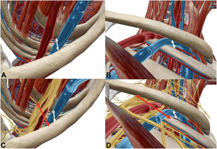
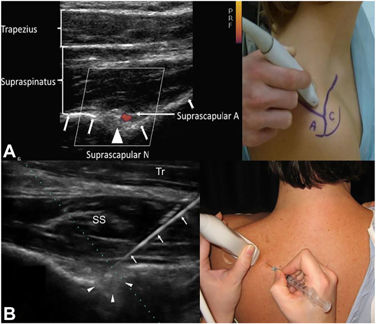
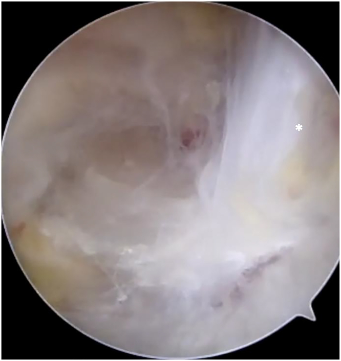
Compressive pathology in the supraclavicular and infraclavicular fossae is broadly termed "thoracic outlet syndrome," with the large majority being neurogenic in nature. These are challenging conditions for patients and physicians and require robust knowledge of thoracic outlet anatomy and scapulothoracic kinematics to elucidate neurogenic versus vascular disorders. The combination of repetitive overhead activity and scapular dyskinesia leads to contracture of the scalene muscles, subclavius, and pectoralis minor, creating a chronically distalized and protracted scapular posture. This decreases the volume of the scalene triangle, costoclavicular space, and retropectoralis minor space, with resultant compression of the brachial plexus causing neurogenic thoracic outlet syndrome. This pathologic cascade leading to neurogenic thoracic outlet syndrome is termed pectoralis minor syndrome when primary symptoms localize to the infraclavicular area. Making the correct diagnosis is challenging and requires the combination of complete history, physical examination, advanced imaging, and ultrasound-guided injections. Most patients improve with nonsurgical treatment incorporating pectoralis minor stretching and periscapular and postural retraining. Surgical decompression of the thoracic outlet is reserved for compliant patients who fail nonsurgical management and respond favorably to targeted injections. In addition to prior exclusively open procedures with supraclavicular, infraclavicular, and/or transaxillary approaches, new minimally invasive and targeted endoscopic techniques have been developed over the past decade. They involve the endoscopic release of the pectoralis minor tendon, with additional suprascapular nerve release, brachial plexus neurolysis, and subclavius and interscalene release depending on the preoperative work-up.

Keywords: Endoscopic brachial plexus neurolysis; Neurogenic thoracic outlet syndrome; Pectoralis minor release; Pectoralis minor syndrome; Suprascapular neuropathy.


Figures

Figure 1
Figure 1
Rendering of the brachial plexus and subclavian/axillary vessels passing through the thoracic outlet in the right shoulder. A Anterior view with subtraction of the deltoid, pectoralis major, trapezius, rotator cuff, and conjoint tendon. B Brachial plexus highlighted at the cord level deep to the pectoralis minor, with subtraction of the vasculature. C Inlet view (looking from superior to inferior) of the thoracic outlet. The subclavian vein courses anterior to the anterior scalene in the costoclavicular space. The brachial plexus and subclavian artery pass between the anterior and middle scalenes. D Outlet view (looking from inferior to superior) of the thoracic outlet. (1) anterior scalene, (2) middle scalene, (3) first rib, (4) coracoid process, (5) pectoralis minor muscle, (6) brachial plexus, (7) subclavian artery, and (8) subclavian vein.
Figure 2
Figure 2
Inlet and outlet renderings illustrating anatomic relationships of the neurovascular bundle and osseous structures. A Inlet view with subtraction of the brachial plexus, demonstrating the proximity of the subclavian vessels between the clavicle and first rib. B Outlet view depicting a similar relationship between the vessels and osseous anatomy. C Inlet view with the brachial plexus. Note the relatively posterior position of the plexus and increased distance between the clavicle and first rib at this location. D Outlet view with the brachial plexus. Again, note the markedly increased distance between the clavicle and first rib at the posterior location of the plexus.
Figure 3
Figure 3
Images illustrating the relationship between the brachial plexus and PM within the retropectoralis minor space. A Anterior view, with the brachial plexus highlighted at the level of the cords. B Outlet view of the retropectoralis minor space, again demonstrating the proximity of the cords to the undersurface of the PM.
Figure 4
Figure 4
Appearance of the scapular protraction in 2 patients. A Posterior view with protraction of the left scapula, creating posterior elevation and prominence of the inferior angle. B Lateral and superior views demonstrating protracted resting position of the left scapula, with prominence of the inferior angle posteriorly.
Figure 5
Figure 5
Rendering of the suprascapular nerve and the transverse scapular ligament with subtraction of the deltoid, trapezius, rotator cuff muscles, brachial plexus, vasculature, and PM. A Anterior view. B Posterior view. C Superior view. Note how the branch innervating the supraspinatus takes a sharp turn medially in the supraspinatus fossa immediately beyond the transverse scapular ligament (black arrow indicates the transverse scapular ligament).
Figure 6
Figure 6
A PM length. Measurement of the distance from the medial aspect of the coracoid to the inferior margin of the fourth rib at the sternocostal junction with the patient upright. B Measurement of the contralateral side performed for comparison.
Figure 7
Figure 7
Large subcoracoid cyst noted on the magnetic resonance imaging of the left shoulder and brachial plexus in a patient with vague, deep pain around the anterior shoulder and upper chest, worse with repetitive activity. A Coronal view; cyst marked with white ∗. B Sagittal view; cyst marked with white ∗. C Anterior coronal slice; cyst marked with white ∗, plexus marked with white ˆ. D Posterior coronal slice; cyst marked with white ∗, coracoid marked with white star, and humeral head marked with white downward arrow. The patient was treated with arthroscopic PM release, cyst decompression, and brachial plexus neurolysis, with resolution of her symptoms and return to activity.
Figure 8
Figure 8
Ultrasound-guided injection of local anesthetic targeted to the PM insertion on the coracoid. The ∗ indicates coracoid, ˆ indicates PM insertion, and white upward arrows indicate needle). PMa, pectoralis major.
Figure 9
Figure 9
Ultrasound-guided injection of local anesthetic targeted to the scalene triangle. The white arrows indicate the needle. AS, anterior scalene; CA, carotid artery; IJ, internal jugular vein; MS, middle scalene; SCM, sternocleidomastoid.
Figure 10
Figure 10
Figure-of-eight brace exerts a constant posterior force on the scapula to combat chronic protraction, augmenting postural retraining.
Figure 11
Figure 11
Ultrasound-guided injection of local anesthetic targeted to the suprascapular notch, demonstrating 2 different injection techniques. A Superior approach, with Doppler use to identify the suprascapular artery (white arrows with split tails demonstrate needle path and infiltration of anesthetic fluid). B Posterior approach. The white arrows indicate the needle, and the white arrowheads indicate the infiltration of fluid in the suprascapular notch. T, trapezius.
Figure 12
Figure 12
Arthroscopic portals for suprascapular neurolysis, PM release, brachial plexus neurolysis, and infraclavicular thoracic outlet decompression (right shoulder). Portals A–D are used for suprascapular neurolysis. Portals A, B, E, F are used for PM release, brachial plexus neurolysis, and infraclavicular thoracic outlet decompression. A, lateral; B, anterolateral; C, transtrapezial; D, medial transtrapezial; E, anterior; F, anteromedial.
Figure 13
Figure 13
Arthroscope views from the lateral portal while the anterolateral working portal is created via needle localization. These are the initial viewing and working portals for both suprascapular nerve release and PM release.
Figure 14
Figure 14
Viewing from the same lateral portal, the base of the coracoid is seen below the electrocautery wand. Medial advancement progresses toward the suprascapular notch. The ∗ indicates the base of the coracoid.
Figure 15
Figure 15
Viewing from the same lateral portal, the vertically oriented fibers of the coracoclavicular ligaments are seen (white ∗) as one advances posteriorly around the coracoid base.
Figure 16
Figure 16
Arthroscopic suprascapular nerve release of the right shoulder. A View from the subacromial space via the lateral portal and 30° arthroscope, progressing medially following the CA ligament and releasing along the anterior border of the supraspinatus muscle until the transverse scapular ligament is encountered posterior to the coracoid. B Needle localization creating the medial transtrapezial working portal. C Arthroscopic scissors introduced through this working portal, releasing the transverse scapular ligament. The suprascapular nerve is safely visualized inferior to the ligament. The suprascapular artery runs anterior to posterior over the ligament, and is displaced posterior-medial to the scissors to ensure it remains protected. D Released suprascapular nerve. The ∗ indicates suprascapular nerve, and ˆ indicates suprascapular artery. TSL, transverse scapular ligament.
Figure 17
Figure 17
Arthroscope now transitioned to the anterolateral portal for viewing. The anterior portal was also created, with a switching stick serving as a retractor in the subdeltoid and subpectoral space. Scalpel is preparing to create the anteromedial working portal following needle localization.
Figure 18
Figure 18
Arthroscopic PM release of the right shoulder. A View from the anterolateral portal with standard 30° arthroscope, demonstrating the coracoacromial ligament (∗) and conjoint tendon (ˆ). B View from the same anterolateral portal with 70° arthroscope, demonstrating the classic “T” appearance of the coracoacromial ligament (∗), conjoint tendon (ˆ), and PM (downward white arrow) converging on the coracoid process. C Release of the PM tendon insertion (downward white arrow) off the medial coracoid using electrocautery. The conjoint tendon is also seen in this view (ˆ). D Continued release of the coracoid with inferior and medial retraction of the PM tendon (downward white arrow). This retraction of the released tendon is routinely noted in patients with PMS and NTOS.
Figure 19
Figure 19
Grasper maneuver to appreciate PM tension after release. A The grasper is used to hold the released lateral edge of the tendon (right side of image). B, C The grasper pulls the tendon laterally, back toward the coracoid insertion. D Immediate inferomedial tendon retraction occurs upon release of the grasper.
Figure 20
Figure 20
Brachial plexus arthroscopic neurolysis following PM release. A PM completely released (downward white arrow) with classic inferomedial retraction. Loose areolar tissue is seen deep, indicating that the retropectoralis minor space is now open. B Release of adhesions and areolar tissue in the retropectoralis minor space uncovers the lateral cord of the brachial plexus. C Continued proximal release uncovers the axillary artery. The medial cord is partially visualized at the bottom right. D Further proximal release presents the subclavius muscle on the inferior surface of the clavicle. This is released with electrocautery, completing the infraclavicular thoracic outlet release. AA, axillary artery; LC, lateral cord; MC, medial cord; S, subclavius muscle).

References

    1. Atroshi I., Gummesson C., Johnsson R., Ornstein E., Ranstam J., Rosén I. Prevalence of carpal tunnel syndrome in a general population. JAMA. 1999;282(2):153–158. - PubMed
    1. An T.W., Evanoff B.A., Boyer M.I., Osei D.A. The prevalence of cubital tunnel syndrome: a cross-sectional study in a U.S. metropolitan cohort. J Bone Joint Surg Am. 2017;99(5):408–416. - PMC - PubMed
    1. Mondelli M., Grippo A., Mariani M., et al. Carpal tunnel syndrome and ulnar neuropathy at the elbow in floor cleaners. Neurophysiol Clin. 2006;36(4):245–253. - PubMed
    1. Jones M.R., Prabhakar A., Viswanath O., et al. Thoracic outlet syndrome: a comprehensive review of pathophysiology, diagnosis, and treatment. Pain Ther. 2019;8(1):5–18. - PMC - PubMed
    1. Kuhn J.E., Lebus V.G.F., Bible J.E. Thoracic outlet syndrome. J Am Acad Orthop Surg. 2015;23(4):222–232. - PubMed

LinkOut - more resources