Review
. 2022 Sep 11;5(4):547-560.
doi: 10.1016/j.jhsg.2022.07.008. eCollection 2023 Jul.

Modern Treatment of Cubital Tunnel Syndrome: Evidence and Controversy

Affiliations
Review

Modern Treatment of Cubital Tunnel Syndrome: Evidence and Controversy

Alexander Graf et al. J Hand Surg Glob Online. .

Abstract

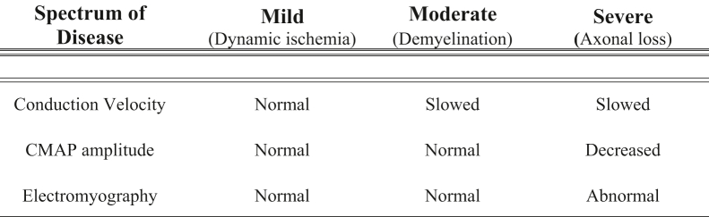
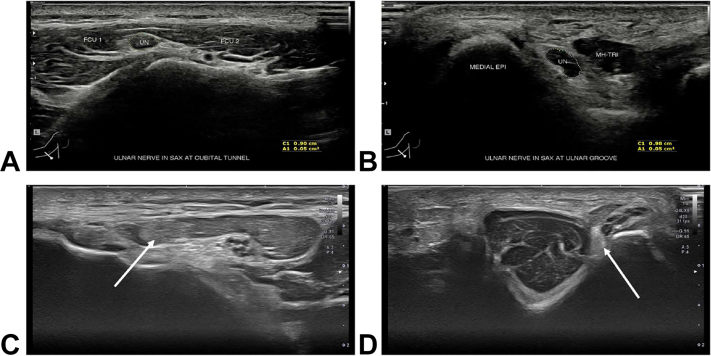
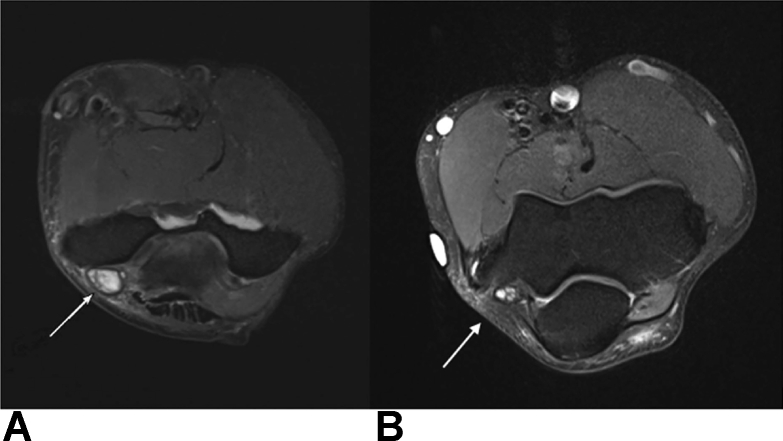
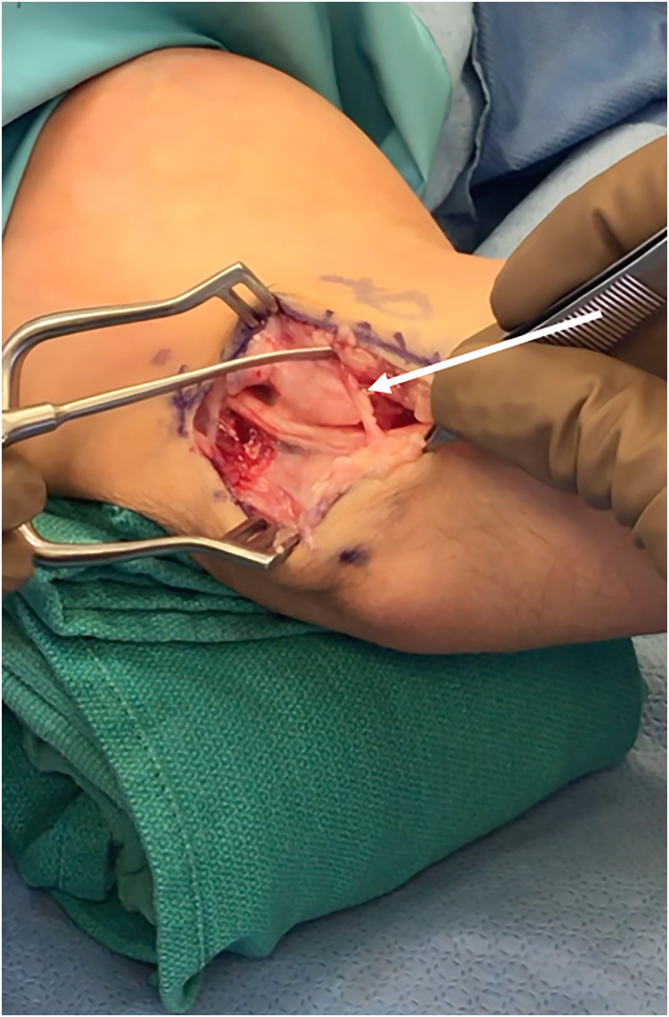
Cubital tunnel syndrome is the second most common peripheral mononeuropathy in the upper extremity. However, the diagnosis and treatment of cubital tunnel syndrome remains controversial without a standard algorithm. Although diagnosis can often be made from the patient's history and physical examination alone, electrodiagnostic studies, ultrasound, computed tomography (CT), and magnetic resonance image (MRI) can also be useful in diagnosing the disease and selecting the most appropriate treatment option. Treatment options include conservative nonoperative techniques as well as various surgical options, including in situ decompression with or without transposition, medial epicondylectomy, and nerve transfer in advanced disease. The purpose of this review is to summarize the most up-to-date literature regarding cubital tunnel syndrome and propose a treatment algorithm to provide clarity about the challenges of treating this complex patient population.

Keywords: Cubital tunnel syndrome; Electrodiagnostic testing; Nerve compression syndrome; Nerve transfer; Ulnar nerve.


Figures

Figure 1
Figure 1
Points of ulnar nerve compression in CuTS.
Figure 2
Figure 2
Intraoperative photograph of right elbow depicting an anomalous anconeus epitrochlearis (arrow) present in a patient with CuTS.
Figure 3
Figure 3
Elbow extension splinting.
Figure 4
Figure 4
Ulnar nerve branching patterns at the elbow and wrist. Used with permission from Andrews et al.
Figure 5
Figure 5
Topography of the ulnar nerve in the forearm and hand from Moore et al. Used with permission from Elsevier.
Figure 6
Figure 6
Intraoperative photograph of the right elbow depicting preservation of blood supply during anterior transposition of the nerve for treatment of CuTS. The arrow indicates the intact posterior recurrent ulnar artery.
Figure 7
Figure 7
Physical examination findings and signs in patients with CuTS. A Ulnar claw hand with atrophy of the FDI. B Wartenberg sign. C Froment sign. D Jeanne sign.
Figure 8
Figure 8
Provocative physical examination maneuvers that are useful in the diagnosis of CuTS. A Tinel sign test. B Flexion compression test. C Scratch collapse test before collapse. D Scratch collapse test after collapse.
Figure 9
Figure 9
High-resolution ultrasound depicting dynamic instability of a hypermobile ulnar nerve (arrow). A The elbow in extension with the ulnar nerve reduced. B The elbow in hyperflexion with the ulnar nerve subluxated and no longer visible.
Figure 10
Figure 10
EDX findings in CuTS.
Figure 11
Figure 11
High-resolution ultrasound for diagnosis in CuTS. A The ulnar nerve (UN) as it courses between the 2 bellies of the FCU. B The UN as it travels around the elbow in the ulnar groove between the medial epicondyle (MEDIAL EPI) of the humerus and the medial head of the triceps (MH-TRI). C The UN (arrows) in a patient which CuTS at the FCU. D The UN (arrows) in a patient with CuTS at the FCU and perched on the medial epicondyle. The nerve’s morphological appearance is abnormally hypoechoic and has lost its normal fascicular echo pattern.
Figure 12
Figure 12
Axial MRI using T2-weighted imaging of the right elbow. A An enlarged and high signal intensity ulnar nerve in a patient with CuTS. B Comparison MRI in a patient without CuTS depicting the internal topography of the ulnar nerve fascicles.
Figure 13
Figure 13
Intraoperative photograph of in situ decompression of the ulnar nerve in a patient with CuTS. The arrow indicates preservation of the medial antebrachial cutaneous nerve.
Figure 14
Figure 14
Ulnar nerve transposition surgery. A Anterior subcutaneous transposition. B Submuscular ulnar nerve transposition.
Figure 15
Figure 15
Submuscular transposition of the ulnar nerve demonstrating both the musculofascial lengthening of the flexor-pronator mass and the distal and proximal flaps of the fasciodermal sling.
Figure 16
Figure 16
Intraoperative photograph of anterior interosseous nerve (AIN)-to-ulnar motor nerve transfer for CuTS. Anterior interosseous nerve (arrow) as it is transferred to the ulnar nerve.
Figure 17
Figure 17
Algorithm for CuTS diagnosis and treatment. ∗Clinical examination consistent with CuTS is defined as having paresthesias in the distribution of the ulnar nerve, symptoms caused by elbow flexion, a positive Tinel sign at the medial elbow, and/or widened 2-point discrimination in ulnar nerve distribution. ∗∗Moderate findings of EDX include decreased conduction velocity. ∗∗∗Severe findings of EDX include decreased CMAP with/without abnormal electromyography findings.

References

    1. Staples J.R., Calfee R. Cubital tunnel syndrome: current concepts. J Am Acad Orthop Surg. 2017;25(10):e215–e224. - PubMed
    1. An T.W., Evanoff B.A., Boyer M.I., Osei D.A. The prevalence of cubital tunnel syndrome: a cross-sectional study in a U.S. metropolitan cohort. J Bone Joint Surg Am. 2017;99(5):408–416. - PMC - PubMed
    1. Naran S., Imbriglia J.E., Bilonick R.A., Taieb A., Wollstein R. A demographic analysis of cubital tunnel syndrome. Ann Plast Surg. 2010;64(2):177–179. - PubMed
    1. Yahya A., Malarkey A.R., Eschbaugh R.L., Bamberger H.B. Trends in the surgical treatment for cubital tunnel syndrome: a survey of members of the American Society for Surgery of the Hand. Hand (N Y) 2018;13(5):516–521. - PMC - PubMed
    1. Soltani A.M., Best M.J., Francis C.S., Allan B.J., Panthaki Z.J. Trends in the surgical treatment of cubital tunnel syndrome: an analysis of the national survey of ambulatory surgery database. J Hand Surg. 2013;38(8):1551–1556. - PubMed

LinkOut - more resources