

Bovine pericardium membrane (TutoPatch) for emergency repair of total corneal melting over an infected corneal graft
- PMID: 37521803
- PMCID: PMC10384563
- DOI: 10.1016/j.ajoc.2023.101885
Bovine pericardium membrane (TutoPatch) for emergency repair of total corneal melting over an infected corneal graft
Abstract
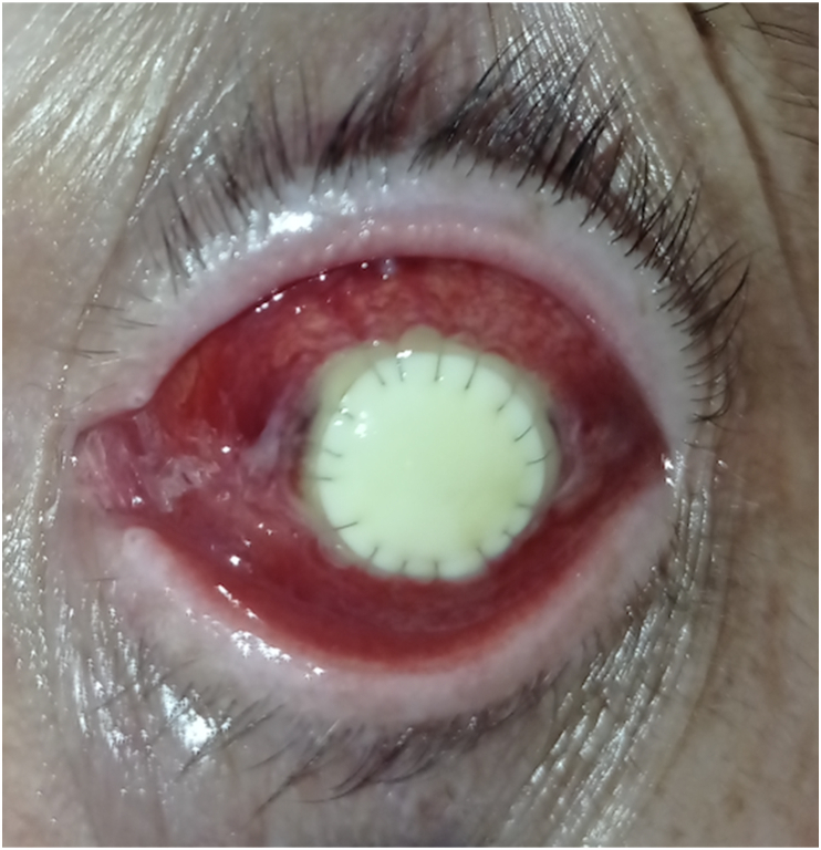
Purpose: Corneal perforation due to severe melting is a very dangerous, sight-threatening condition requiring immediate management due to the high risk of endophthalmitis and critical hypotony. In the case of perforated corneal grafts, retransplantation is usually postponed to avoid the detrimental effects of inflammation on the new graft. We describe the first case of the use of a TutoPatch graft for emergency replacement of a lamellar graft perforation over acute infectious total melting.
Observations: A 42-year-old male patient presented to the Emergency Department with pain in the left eye, which was red photophobic. He had been treated with bilateral deep anterior lamellar keratoplasty (DALK) for advanced keratoconus 5 years previously and had been experiencing recurrent corneal ulcers in the left eye within the last 8 months. Clinical examination documented corneal perforation over acute infectious melting involving the total graft surface in the left eye. The infected graft was removed along with the perforated infected residual Descemet membrane, and a double-layer TutoPatch covering was sutured to the host's margin with 10.0 nylon. The covering was left in place for three weeks, allowing the patient to undergo retransplant three weeks later without complications.
Conclusions and importance: TutoPatch covering can be safely used as an easy-to-preserve emergency material for a temporary bridge to retransplantation in large acute infectious corneal melting.
Keywords: Bacterial keratitis; Corneal graft; Corneal melting; Corneal perforation; DALK; TutoPatch.
© 2023 Published by Elsevier Inc.
Conflict of interest statement
The authors declare that they have no known competing financial interests or personal relationships that could have appeared to influence the work reported in this paper.
Figures


Similar articles
-
Corneal Perforation as a Complication of Fungal Interface Infectious Keratitis after Deep Anterior Lamellar Keratoplasty.Middle East Afr J Ophthalmol. 2021 Dec 31;28(3):184-188. doi: 10.4103/meajo.meajo_114_21. eCollection 2021 Jul-Sep. Middle East Afr J Ophthalmol. 2021. PMID: 35125802 Free PMC article.
-
Bovine pericardium membrane (tutopatch) combined with solid platelet-rich plasma for the management of perforated corneal ulcers.Cornea. 2013 May;32(5):619-24. doi: 10.1097/ICO.0b013e31825a6d9a. Cornea. 2013. PMID: 22929158
-
Use of bovine pericardium (Tutopatch®) graft for surgical repair of deep melting corneal ulcers in dogs and corneal sequestra in cats.Vet Ophthalmol. 2014 Mar;17(2):91-9. doi: 10.1111/vop.12047. Epub 2013 Apr 28. Vet Ophthalmol. 2014. PMID: 23621151
-
Infectious interface keratitis (IIK) following lamellar keratoplasty: A literature review.Ocul Surf. 2019 Oct;17(4):635-643. doi: 10.1016/j.jtos.2019.08.001. Epub 2019 Aug 12. Ocul Surf. 2019. PMID: 31415815
-
[Partial visual rehabilitation 5 and 6 years after a Gundersen total conjunctival flap procedure].Ophthalmologe. 2022 Feb;119(2):203-208. doi: 10.1007/s00347-021-01503-4. Epub 2021 Sep 28. Ophthalmologe. 2022. PMID: 34581853 Review. German.
References
Publication types
LinkOut - more resources
Full Text Sources
