. 2023 Jul 13:11:1222762.
doi: 10.3389/fpubh.2023.1222762. eCollection 2023.

Effects of ozone exposure on lipid metabolism in Huh-7 human hepatoma cells

Affiliations

Effects of ozone exposure on lipid metabolism in Huh-7 human hepatoma cells

Jianhao Peng et al. Front Public Health. .

Abstract

Ozone pollution is a major environmental concern. According to recent epidemiological studies, ozone exposure increases the risk of metabolic liver disease. However, studies on the mechanisms underlying the effects of ozone exposure on hepatic oxidative damage, lipid synthesis, and catabolism are limited. In this study, Huh-7 human hepatocellular carcinoma cells were randomly divided into five groups and exposed to 200 ppb O3 for 0, 1, 2, 4, and 8 h. We measured the levels of oxidative stress and analyzed the changes in molecules related to lipid metabolism. The levels of oxidative stress were found to be significantly elevated in Huh-7 hepatocellular carcinoma cells after O3 exposure. Moreover, the expression levels of intracellular lipid synthases, including SREBP1, FASN, SCD1, and ACC1, were enhanced. Lipolytic enzymes, including ATGL and HSL, and the mitochondrial fatty acid oxidase, CPT1α, were inhibited after O3 exposure. In addition, short O3 exposure enhanced the expression of the intracellular peroxisomal fatty acid β-oxidase, ACOX1; however, its expression decreased adaptively with longer exposure times. Overall, O3 exposure induces an increase in intracellular oxidative stress and disrupts the normal metabolism of lipids in hepatocytes, leading to intracellular lipid accumulation.

Keywords: Huh-7; O3; ROS; lipid metabolism; oxidative stress.


Conflict of interest statement

The authors declare that the research was conducted in the absence of any commercial or financial relationships that could be construed as a potential conflict of interest.

Figures

Figure 1
Figure 1
Results of cell viability assay of Huh-7 cells after O3 exposure. (A) Results of Trypan blue staining in the background with different time gradients of ozone exposure. (B) Huh-7 cells were treated with control or 200 ppb O3 for 1, 2, 4, and 8 h, respectively, and cell viability was measured using CCK-8 method. *p < 0.05, **p < 0.01,***p < 0.001 vs. control group.
Figure 2
Figure 2
CM-H2DCFDA staining results of Huh-7 cells after 200 ppb O3 exposure. (A) Intracellular ROS content after 200 ppb O3 exposure for 0 h. (B) Intracellular ROS content after 200 ppb O3 exposure for 1 h. (C) Intracellular ROS content after 200 ppb O3 exposure for 2 h. (D) Intracellular ROS content after 200 ppb O3 exposure for 4 h. (E) Intracellular ROS content after 200 ppb O3 exposure for 8 h.
Figure 3
Figure 3
Effect of O3 exposure on protein expression of oxidative stress-related enzymes. Protein expression levels of CYP1A1 (A) and SOD1 (B) in Huh-07 cells at different time gradients (0, 1, 2, 4, and 8 h) after O3 exposure are expressed as mean ± SE (n = 3 cells/group). *p < 0.05, **p < 0.01,***p < 0.001 vs. control group.
Figure 4
Figure 4
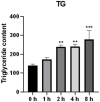
Effect of O3 exposure on intracellular triglyceride content. The level of TG in Huh-07 cells at different time gradients (0, 1, 2, 4, and 8 h) after O3 exposure are expressed as mean ± SE (n = 3 cells/group). *p < 0.05, **p < 0.01,***p < 0.001 vs. control group.
Figure 5
Figure 5
Effect of O3 exposure on mRNA expression of lipid synthesis-related enzymes. mRNA expression levels of FASN (A), SCD1 (B), SREBP1 (C), and ACC1 (D) in Huh-7 cells at different time gradients (0, 1, 2, 4, and 8 h) after O3 exposure are expressed as mean ± SE (n = 3 cells/group). *p < 0.05, **p < 0.01 vs. control group.
Figure 6
Figure 6
Effect of O3 exposure on mRNA expression of genes encoding key enzymes or regulators involved in fatty acid oxidation and lipolysis in Huh-7 cells. mRNA expression levels of PPARα (A), CPT1α (B), ACOX1 (C), ATGL (D), and HSL (E) in Huh-7 cells at different time gradients (0, 1, 2, 4, and 8 h) after O3 exposure are expressed as mean ± SE (n = 3 cells/group). *p < 0.05, **p < 0.01,***p < 0.001 vs. control group.
Figure 7
Figure 7
Effects of O3 exposure on the protein expression of lipid metabolism-related genes in Huh-7 cells at different time gradients (0, 1, 2, 4, and 8 h). (A) Protein expression of FASN; (B) Protein expression of SCD1; (C) Protein expression of SREBP1; (D) Protein expression of ACC1; (E) Protein expression of PPARα; (F) Protein expression of CPT1α; (G) Protein expression of ACOX1; (H) Protein expression of ATGL; (I) Protein expression of HSL. Data were expressed as mean ± SE (n = 3 cells/group). *p < 0.05, **p < 0.01,***p < 0.001 vs. control group.

References

    1. IHME State of global air 2020:a special report on Globle exposure to air pollution and its health impacts. IHME. (2020).
    1. Landrigan PJ, Fuller R, Acosta NJR, Adeyi O, Arnold R, Basu NN, et al. . The lancet commission on pollution and health. Lancet. (2018) 391:462–512. doi: 10.1016/S0140-6736(17)32345-0 - DOI - PubMed
    1. Lu F, Xu D, Cheng Y, Dong S, Guo C, Jiang X, et al. . Systematic review and meta-analysis of the adverse health effects of ambient PM2.5 and PM10 pollution in the Chinese population. Environ Res. (2015) 136:196–204. doi: 10.1016/j.envres.2014.06.029, PMID: - DOI - PubMed
    1. Bocci V, Valacchi G, Corradeschi F, Aldinucci C, Silvestri S, Paccagnini E, et al. . Studies on the biological effects of ozone: 7. Generation of reactive oxygen species (ROS) after exposure of human blood to ozone. J Biol Regul Homeost Agents. (1998) 12:67–75. PMID: - PubMed
    1. Tian L, Li N, Li K, Tan Y, Han J, Lin B, et al. . Ambient ozone exposure induces ROS related-mitophagy and pyroptosis via NLRP3 inflammasome activation in rat lung cells. Ecotoxicol Environ Saf. (2022) 240:113663. doi: 10.1016/j.ecoenv.2022.113663, PMID: - DOI - PubMed

Publication types