. 2023 Aug;17(4):869-891.
doi: 10.1007/s11571-022-09869-0. Epub 2022 Aug 30.

Multiscale entropy of ADHD children during resting state condition

Affiliations

Multiscale entropy of ADHD children during resting state condition

Brenda Y Angulo-Ruiz et al. Cogn Neurodyn. 2023 Aug.

Abstract

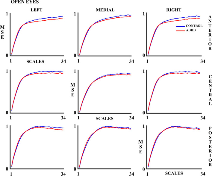
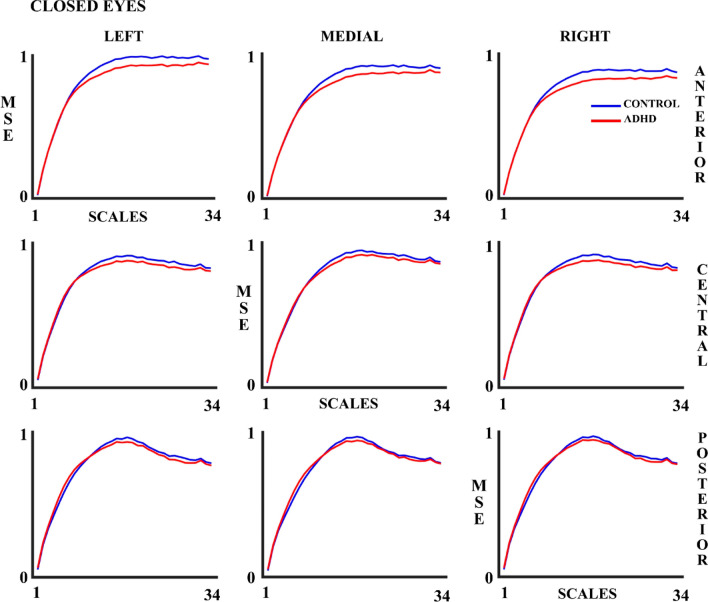
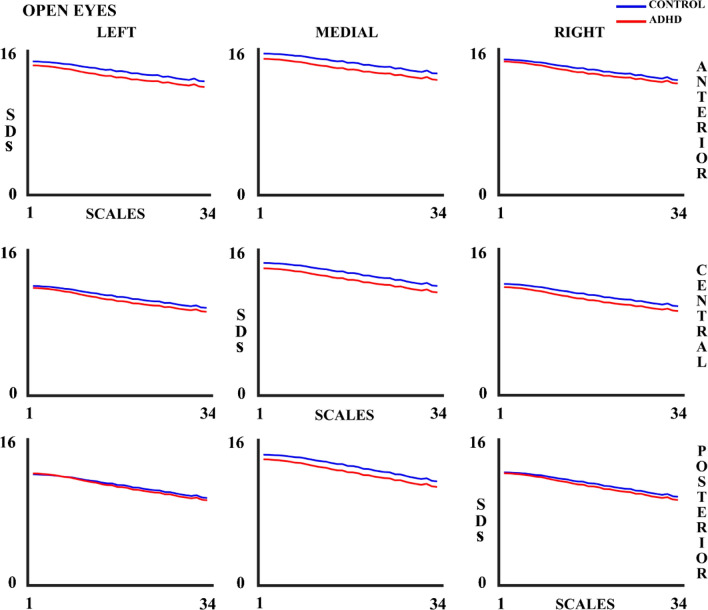
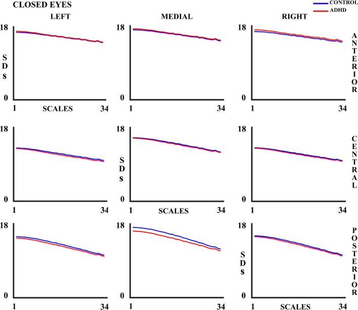
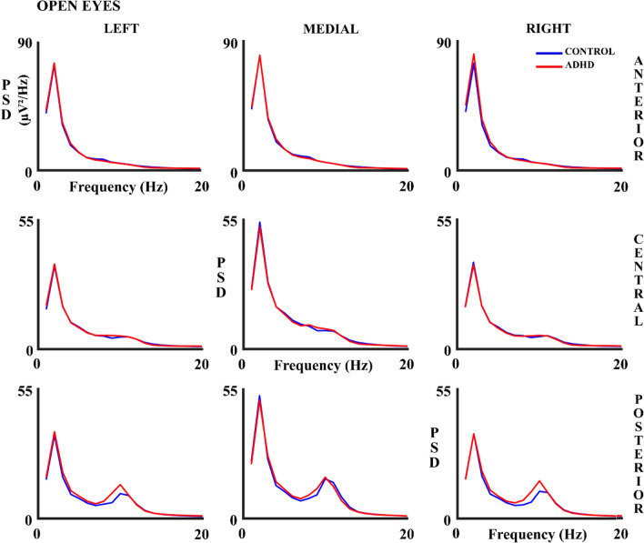
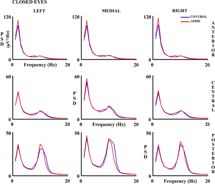
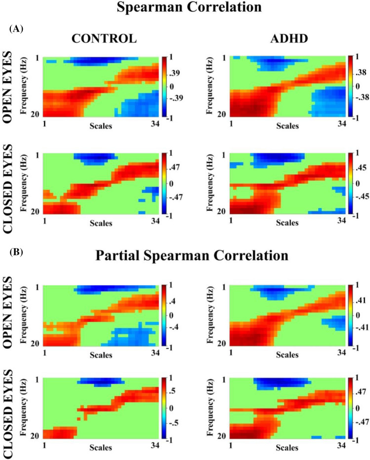
This present study aims to investigate neural mechanisms underlying ADHD compared to healthy children through the analysis of the complexity and the variability of the EEG brain signal using multiscale entropy (MSE), EEG signal standard deviation (SDs), as well as the mean, standard deviation (SDp) and coefficient of variation (CV) of absolute spectral power (PSD). For this purpose, a sample of children diagnosed with attention-deficit/hyperactivity disorder (ADHD) between 6 and 17 years old were selected based on the number of trials and diagnostic agreement, 32 for the open-eyes (OE) experimental condition and 25 children for the close-eyes (CE) experimental condition. Healthy control subjects were age- and gender-matched with the ADHD group. The MSE and SDs of resting-state EEG activity were calculated on 34 time scales using a coarse-grained procedure. In addition, the PSD was averaged in delta, theta, alpha, and beta frequency bands, and its mean, SDp, and CV were calculated. The results show that the MSE changes with age during development, increases as the number of scales increases and has a higher amplitude in controls than in ADHD. The absolute PSD results show CV differences between subjects in low and beta frequency bands, with higher variability values in the ADHD group. All these results suggest an increased EEG variability and reduced complexity in ADHD compared to controls.

Supplementary information: The online version contains supplementary material available at 10.1007/s11571-022-09869-0.

Keywords: ADHD; Complexity metrics; Multiscale entropy; Power spectral density; Resting-state; Variability.


Figures

Fig. 1
Fig. 1
Localization and collapse of electrodes by regions. The colors indicate the nine defined scalp areas for electrodes collapse. The 30 electrodes are divided into two spatial dimensions (lateral and anterior–posterior) which have three values each one: left, middle, and right; and anterior, central, and posterior, respectively
Fig. 2
Fig. 2
Multiscale Entropy (MSE) for 34 scales in control and ADHD subjects in the open eyes (OE) experimental condition in all the 9 considered areas. The blue line represents the control group and the red line the ADHD group
Fig. 3
Fig. 3
Multiscale Entropy (MSE) for 34 scales in control and ADHD subjects in the closed eyes (CE) experimental condition in all the 9 considered areas. The blue line represents the control group and the red line the ADHD group
Fig. 4
Fig. 4
Standard Deviation (SDs) for 34 scales in control and ADHD subjects in the open eyes (OE) experimental condition in all the 9 considered areas. The blue line represents the control group and the red line the ADHD group
Fig. 5
Fig. 5
Standard Deviation (SDs) for 34 scales in control and ADHD subjects in the closed eyes (CE) experimental condition in all the 9 considered areas. The blue line represents the control group and the red line the ADHD group
Fig. 6
Fig. 6
Absolute Power Spectral Density (PSD) in Hz in control and ADHD subjects for the open eyes (OE) experimental condition in all 9 considered areas. The blue line represents the control group and the red line the ADHD group
Fig. 7
Fig. 7
Absolute Power Spectral Density (PSD) in control and ADHD subjects for the closed eyes (CE) experimental condition in all 9 considered areas. The blue line represents the control group and the red line the ADHD group
Fig. 8
Fig. 8
A Spearman Correlation between MSE (34 scales) and relative PSD (1–20 Hz) in control and ADHD subjects for both experimental conditions B Partial Spearman correlation, controlling for age (in days) in open and closed eye conditions for both groups of subjects. The cutoff value for each correlation, being significant when corrected by FDR, is indicated in the graphs. The red color represents positive correlations and the blue one represents negative correlations

Similar articles

Cited by

References

    1. Angulo-Ruiz BY, Muñoz V, Rodríguez-Martínez EI, Gómez C. Absolute and relative variability changes of the resting state brain rhythms from childhood and adolescence to young adulthood. Neurosci Lett. 2021;749:135747. doi: 10.1016/j.neulet.2021.135747. - DOI - PubMed
    1. Arns M, Conners CK, Kraemer HC. A decade of EEG Theta/Beta ratio research in ADHD. J Atten Disord. 2013;17(5):374–383. doi: 10.1177/1087054712460087. - DOI - PubMed
    1. Barry RJ, Clarke AR, Johnstone SJ. A review of electrophysiology inattention-deficit/hyperactivity disorder: I qualitative and quantitative electroencephalography. Clin Neurophysiol. 2003;114(2):171–183. doi: 10.1016/s1388-2457(02)00362-0. - DOI - PubMed
    1. Barry RJ, Clarke AR, Johnstone SJ, McCarthy R, Selikowitz M. Electroencephalogram /β ratio and arousal in attention-deficit/hyperactivity disorder: evidence of independent processes. Biol Psychiatry. 2009;66(4):398–401. doi: 10.1016/j.biopsych.2009.04.027. - DOI - PubMed
    1. Benjamini Y, Hochberg Y. Controlling the false discovery rate: a practical and powerful approach to multiple testing. J R Stat Soc. 1995;57(1):289–300. doi: 10.1111/j.2517-6161.1995.tb02031.x. - DOI

LinkOut - more resources