. 2023 Oct 4;32(20):2966-2980.
doi: 10.1093/hmg/ddad122.

Human herpesvirus 8 ORF57 protein is able to reduce TDP-43 pathology: network analysis identifies interacting pathways

Affiliations

Human herpesvirus 8 ORF57 protein is able to reduce TDP-43 pathology: network analysis identifies interacting pathways

Chelsea J Webber et al. Hum Mol Genet. .

Abstract

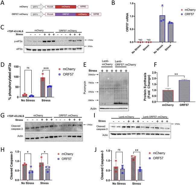
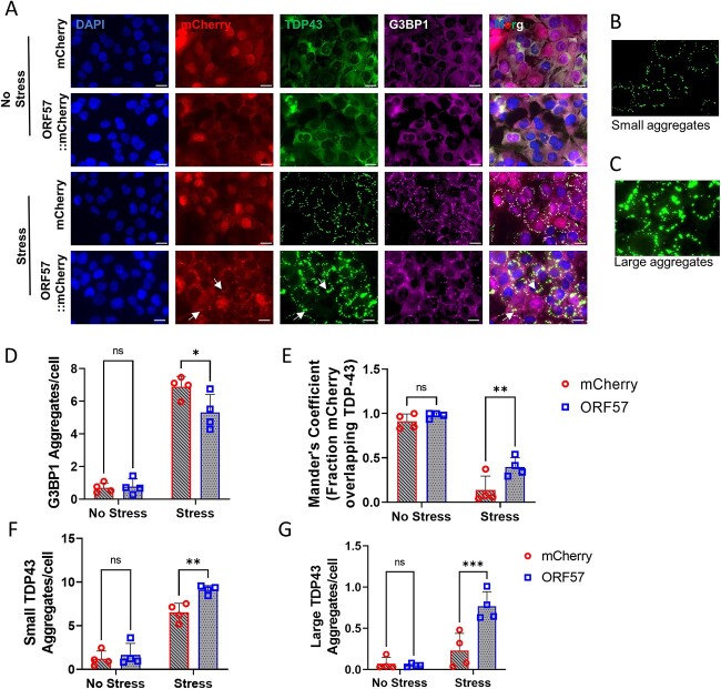
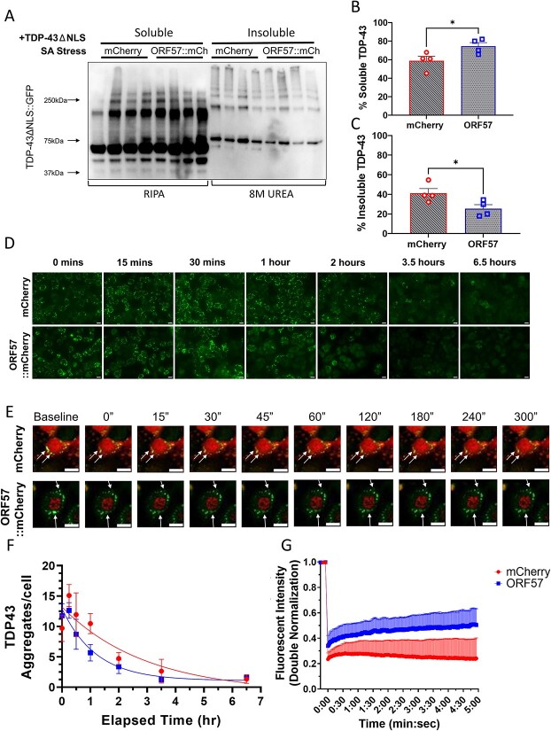
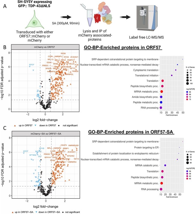
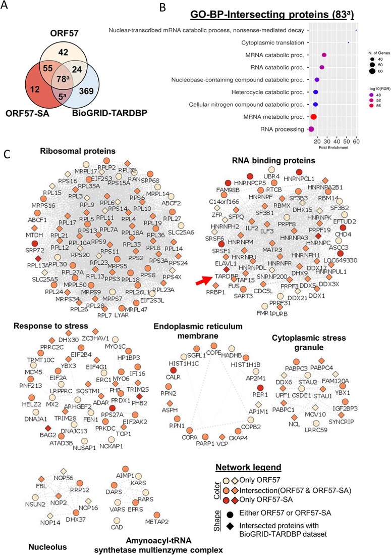
Aggregation of TAR DNA-binding protein 43 kDa (TDP-43) is thought to drive the pathophysiology of amyotrophic lateral sclerosis and some frontotemporal dementias. TDP-43 is normally a nuclear protein that in neurons translocates to the cytoplasm and can form insoluble aggregates upon activation of the integrated stress response (ISR). Viruses evolved to control the ISR. In the case of Herpesvirus 8, the protein ORF57 acts to bind protein kinase R, inhibit phosphorylation of eIF2α and reduce activation of the ISR. We hypothesized that ORF57 might also possess the ability to inhibit aggregation of TDP-43. ORF57 was expressed in the neuronal SH-SY5Y line and its effects on TDP-43 aggregation characterized. We report that ORF57 inhibits TDP-43 aggregation by 55% and elicits a 2.45-fold increase in the rate of dispersion of existing TDP-43 granules. These changes were associated with a 50% decrease in cell death. Proteomic studies were carried out to identify the protein interaction network of ORF57. We observed that ORF57 directly binds to TDP-43 as well as interacts with many components of the ISR, including elements of the proteostasis machinery known to reduce TDP-43 aggregation. We propose that viral proteins designed to inhibit a chronic ISR can be engineered to remove aggregated proteins and dampen a chronic ISR.


Figures

Figure 1
Figure 1
ORF57 protects against oxidative stress in TDP-43𑀏NLS overexpressing SH-SY5Y cells. Experiments conducted in mCherry and ORF57 mCherry express ing SH-SY5Y TDP-43ΔNLS cells with and without SA stress. (A) Constructs for mCherry and ORF57::mCherry lentivirus. (B) Overexpression of ORF57 in SH-SY5Y cells by rt-qPCR. (C) Western blot for phosphorylated eIF2α and total eIF2α. (D) Quantification of C. (E) SUnSET of mCherry and ORF57 expressing TDP-43ΔNLS cells with SA stress. (F) Quantification of E. (G) Western blot of cleaved caspase-3 in TDP-43ΔNLS cells. (H) Quantification of G. (I) Western blot of cleaved caspase-3 in non-overexpressing SH-SY5Y cells. (J) Quantification of I.
Figure 2
Figure 2
Stress granule assembly and TDP-43 aggregation dysregulated by ORF57. (A) Immunocytochemistry in SH-SY5Y TDP-43ΔNLS. DAPI (405 nm). mCherry or ORF57::mCherry (594 nm). TDP-43ΔNLS::GFP (488 nm). G3BP1 (647 nm). Merge, overlay of all images. White arrows indicate overlap between ORF57 and TDP-43 aggregates. (B) Representation of small TDP-43ΔNLS aggregates 10–200 Aggrecount units. (C) Representation of large TDP-43ΔNLS aggregates 200–400 Aggrecount units. (D) Quantification of G3BP1 aggregates in each cell condition from A. (E) Mander’s coefficient of fraction of mCherry overlapping with TDP-43 signal. (F) Quantification of small TDP-43ΔNLS aggregates. (G) Quantification of large TDP-43ΔNLS aggregates. Two-way ANOVA with Bonferroni post hoc comparison, *P < 0.05, **P < 0.01, ***P < 0.0001.
Figure 3
Figure 3
TDP-43 aggregates are more soluble with ORF57. Experiments conducted in mCherry and ORF57 mCherry expressing SH-SY5Y TDP-43ΔNLS cells with SA stress. (A) Western blot of TDP-43ΔNLS::GFP RIPA-soluble and in-soluble fractions. (B) Quantification of %RIPA-Soluble TDP-43ΔNLS::GFP calculated as soluble TDP-43 divided by the sum of soluble and insoluble TDP-43. (C) Quantification of %RIPA-insoluble TDP-43ΔNLS::GFP. (D) Time course experiment measuring TDP-43ΔNLS::GFP aggregate recovery at 15, 30 min, 1 , 2 , 3.5 and 6.5 h after SA removal. (E) Imaging of TDP-43ΔNLS using FRAP. (F) Quantification of D with exponential one-phase decay best fit curves. (G) Analysis of TDP-43ΔNLS mobility using FRAP. mCherry controls cells had a mobile fraction of 0.03 and a t1/2 of 111 s; ORF57::mCherry expressing cells had a higher mobile fraction of 0.2 and a t1/2 of 68 s. Unpaired t-tests, *P < 0.05. Scale bars 20 μm.
Figure 4
Figure 4
ORF57 interactome. (A) Outline of proteomic approach using SH-SY5Y cells expressing GFP::TDP-43ΔNLS. (B) High-confidence ORF57-bound proteins observed under basal conditions and the GO-BP functional annotations terms associated with these enriched proteins. Functional annotation terms were determined using ShinyGO Version 0.76.3 (81) (C) High-confidence ORF57-bound proteins observed in presence of SA and the GO-BP functional annotations terms associated with these enriched proteins. Enriched proteins in basal and stress conditions were considered significant when they have a Log2 fold-change > 0.58 and an FDR adjusted P-value < 0.05.
Figure 5
Figure 5
ORF57 interacts with the TDP-43 protein network. (A) Venn diagram using ORF57, ORF57-SA and the TDP-43 BioGRID-TARDBP) datasets. (B) Functional annotations terms for overlapping proteins between ORF57-SA and BioGRID-TARDBP datasets. (C) Multi-module representation of ORF57-bound protein networks (Using Cytoscape, STRING-db and ClusterMaker2. Node title describes gene names, node shape describes whether the protein was found in either ORF57 datasets (circle) or BioGRID-TARDBP (diamond), and color indicates whether the gene was only found in either ORF57, ORF57-SA or overlapped between them.

References

    1. Neumann, M., Sampathu, D.M., Kwong, L.K., Truax, A.C., Micsenyi, M.C., Chou, T.T., Bruce, J., Schuck, T., Grossman, M., Clark, C.M. et al. (2006) Ubiquitinated TDP-43 in frontotemporal lobar degeneration and amyotrophic lateral sclerosis. Science, 314, 130–133. - PubMed
    1. Igaz, L.M., Kwong, L.K., Xu, Y., Truax, A.C., Uryu, K., Neumann, M., Clark, C.M., Elman, L.B., Miller, B.L., Grossman, M. et al. (2008) Enrichment of C-terminal fragments in TAR DNA-binding protein-43 cytoplasmic inclusions in brain but not in spinal cord of frontotemporal lobar degeneration and amyotrophic lateral sclerosis. Am. J. Pathol., 173, 182–194. - PMC - PubMed
    1. Chou, C.C., Zhang, Y., Umoh, M.E., Vaughan, S.W., Lorenzini, I., Liu, F., Sayegh, M., Donlin-Asp, P.G., Chen, Y.H., Duong, D.M. et al. (2018) TDP-43 pathology disrupts nuclear pore complexes and nucleocytoplasmic transport in ALS/FTD. Nat. Neurosci., 21, 228–239. - PMC - PubMed
    1. Li, Y.R., King, O.D., Shorter, J. and Gitler, A.D. (2013) Stress granules as crucibles of ALS pathogenesis. J. Cell Biol., 201, 361–372. - PMC - PubMed
    1. Gasset-Rosa, F., Lu, S., Yu, H., Chen, C., Melamed, Z., Guo, L., Shorter, J., Da Cruz, S. and Cleveland, D.W. (2019) Cytoplasmic TDP-43 de-mixing independent of stress granules drives inhibition of nuclear import, loss of nuclear TDP-43, and cell death. Neuron, 102, 339–357.e7. - PMC - PubMed

Publication types

MeSH terms

Substances