. 2023 Sep 15;83(18):3115-3130.
doi: 10.1158/0008-5472.CAN-23-0157.

Plasma Membrane Channel TRPM4 Mediates Immunogenic Therapy-Induced Necrosis

Affiliations

Plasma Membrane Channel TRPM4 Mediates Immunogenic Therapy-Induced Necrosis

Santanu Ghosh et al. Cancer Res. .

Abstract

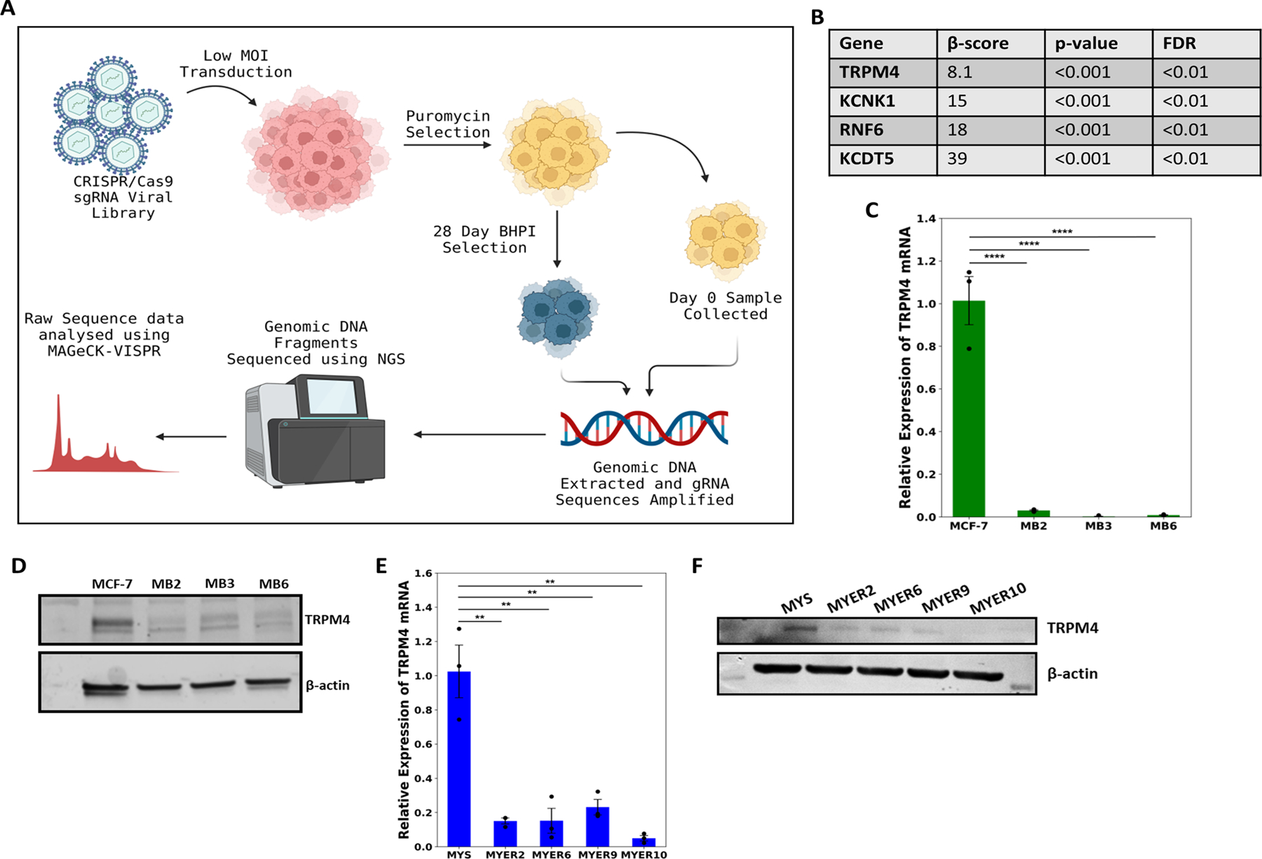
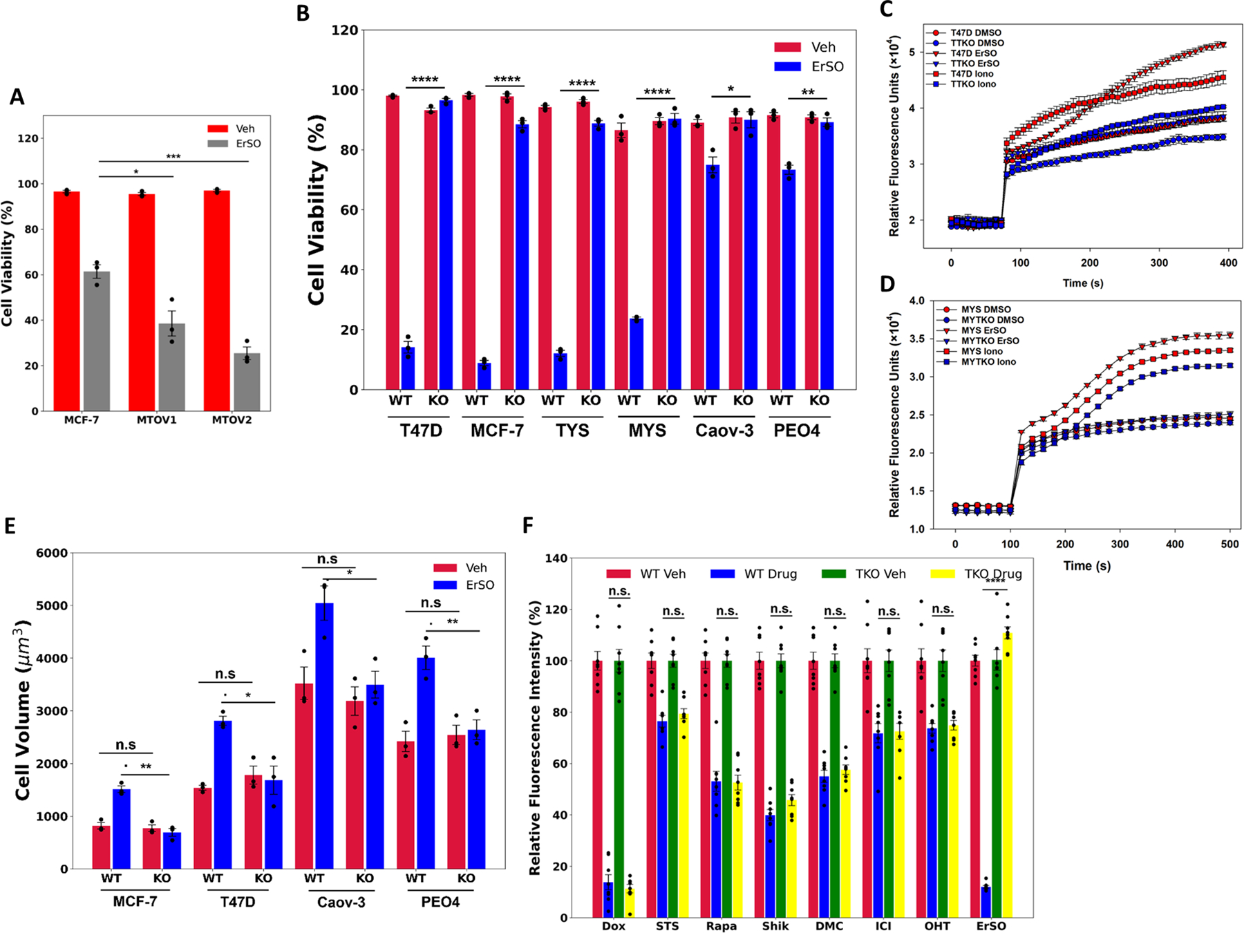
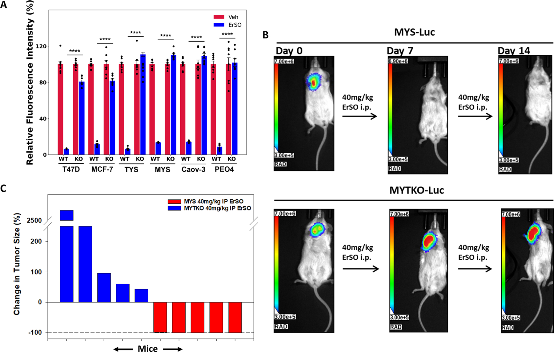
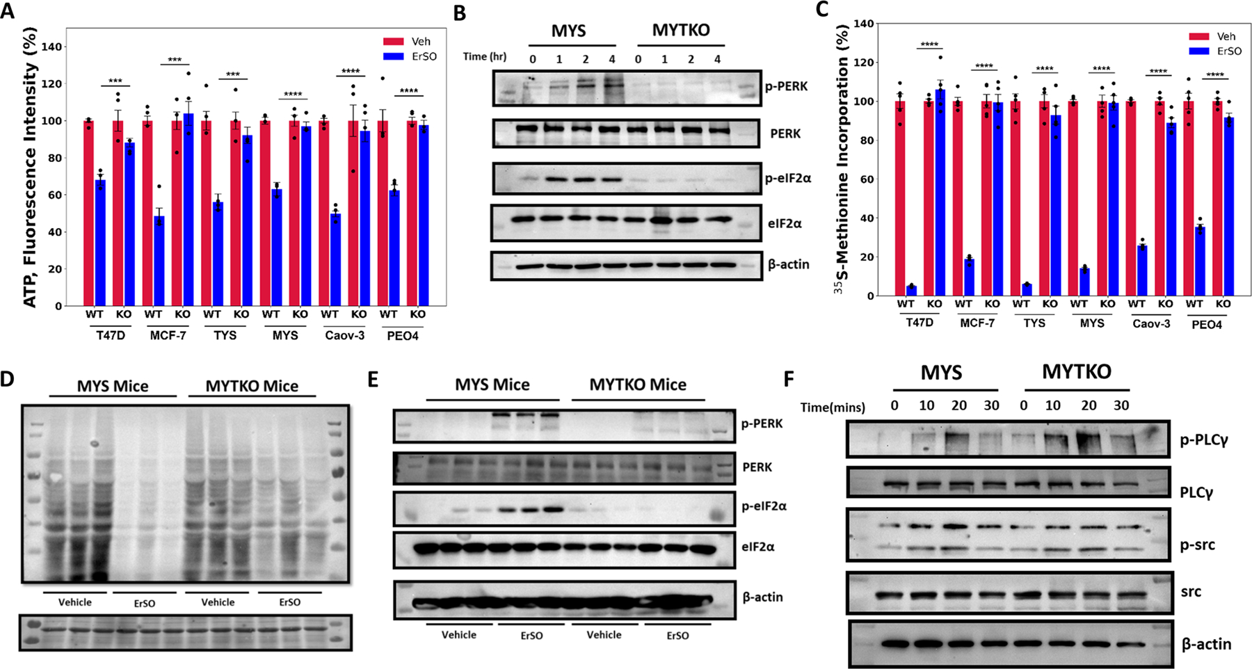
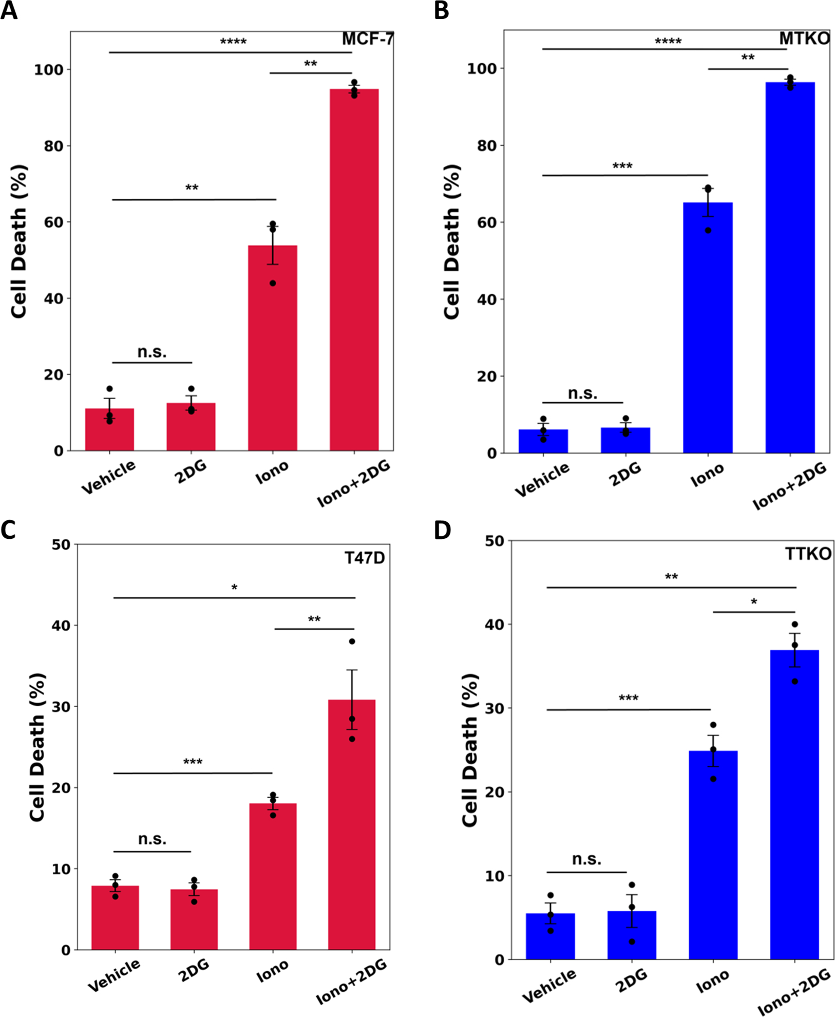
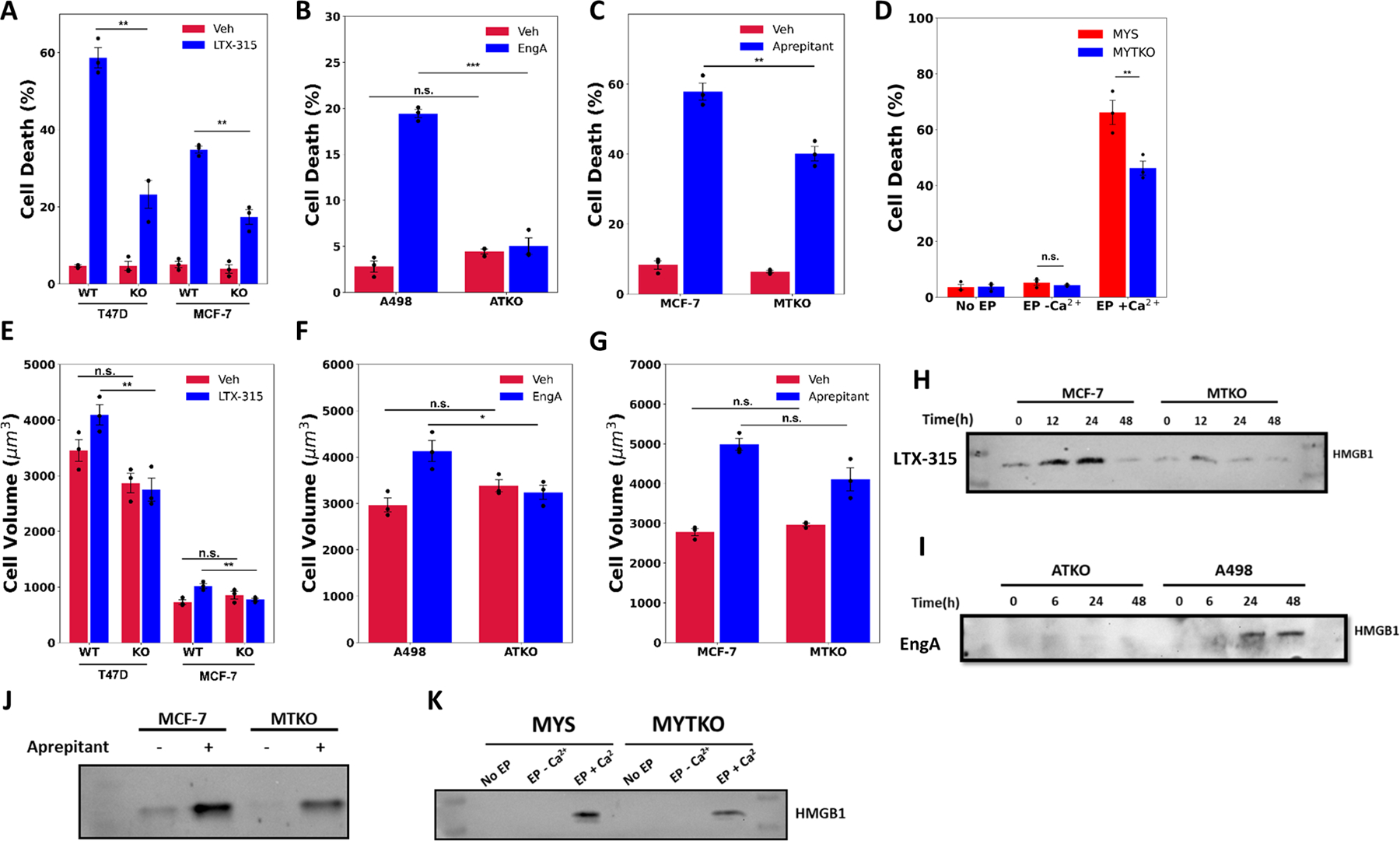
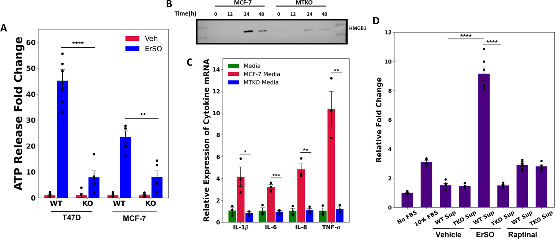
Several emerging therapies kill cancer cells primarily by inducing necrosis. As necrosis activates immune cells, potentially, uncovering the molecular drivers of anticancer therapy-induced necrosis could reveal approaches for enhancing immunotherapy efficacy. To identify necrosis-associated genes, we performed a genome-wide CRISPR-Cas9 screen with negative selection against necrosis-inducing preclinical agents BHPI and conducted follow-on experiments with ErSO. The screen identified transient receptor potential melastatin member 4 (TRPM4), a calcium-activated, ATP-inhibited, sodium-selective plasma membrane channel. Cancer cells selected for resistance to BHPI and ErSO exhibited robust TRPM4 downregulation, and TRPM4 reexpression restored sensitivity to ErSO. Notably, TRPM4 knockout (TKO) abolished ErSO-induced regression of breast tumors in mice. Supporting a broad role for TRPM4 in necrosis, knockout of TRPM4 reversed cell death induced by four additional diverse necrosis-inducing cancer therapies. ErSO induced anticipatory unfolded protein response (a-UPR) hyperactivation, long-term necrotic cell death, and release of damage-associated molecular patterns that activated macrophages and increased monocyte migration, all of which was abolished by TKO. Furthermore, loss of TRPM4 suppressed the ErSO-induced increase in cell volume and depletion of ATP. These data suggest that ErSO triggers initial activation of the a-UPR but that it is TRPM4-mediated sodium influx and cell swelling, resulting in osmotic stress, which sustains and propagates lethal a-UPR hyperactivation. Thus, TRPM4 plays a pivotal role in sustaining lethal a-UPR hyperactivation that mediates the anticancer activity of diverse necrosis-inducing therapies.

Significance: A genome-wide CRISPR screen reveals a pivotal role for TRPM4 in cell death and immune activation following treatment with diverse necrosis-inducing anticancer therapies, which could facilitate development of necrosis-based cancer immunotherapies.


Conflict of interest statement

Conflict of Interest Disclosure Statement

The Univ. of Illinois has filed patent applications on some compounds described here on which DJS, MWB and PJH are co-inventors (US 20200190029; U.S. Patent Application No. 63/055,583). Some compounds described herein have been licensed to Systems Oncology. PJH is a consultant for Systems Oncology and is on the Systems Oncology SAB.

Figures

Figure 1.
Figure 1.
TRPM4 is important for BHPI and ErSO-induced cell death. A Schematic diagram of the CRISPR/Cas9-based genome-wide negative selection screen performed using BHPI on T47D cells. B Partial list of targets obtained from the genome-wide negative selection screen with BHPI. C Relative expression levels of TRPM4 mRNA in wild type MCF-7 and BHPI-resistant MCF-7 cells (MB) (n=3). D Western blot analysis of TRPM4 protein levels in wild type MCF-7 and MB cells. E Relative expression levels of TRPM4 mRNA in MYS (MCF7-ERαY537S) cells and ErSO-resistant MYS cells (MYER) (n=3). F Western blot analysis of TRPM4 protein levels in MYS cells and MYER cells. All data are mean ± s.e.m. *p<0.05, **p<0.01, ***p<0.001, ****p<0.0001.
Figure 2.
Figure 2.
TRPM4 knockout completely blocks ErSO-induced cell death. Automated Trypan Blue dye exclusion assays comparing A wildtype MCF-7 cells and two TRPM4 overexpressing clonal pools (MTOV1 and MTOV2) treated with either vehicle or 50 nM ErSO for 24 hours (n=3), B ERα positive breast cancer cell lines, T47D, MCF-7, TYS and MYS, ovarian cancer cell lines Caov-3 and PEO4, and their TRPM4 knockout clones (TTKO, MTKO, YTKO, MYTKO, CTKO and PTKO respectively) were treated with either vehicle or ErSO (100 nM for T47D, TTKO, MCF-7, MTKO, TYS, and YTKO; 1 μM for MYS, MYTKO, Caov-3, CTKO, PEO4 and PTKO) for 24 hours (n=3). C-D Sodium influx assay comparing D wild type and TRPM4 knockout T47D cells (TTKO) cells, and E MYS and MYTKO cells treated with vehicle, 10 μM ErSO, or the ionophore ionomycin (10 μM) (n=4). E Cell volume of ERα positive breast cancer cell lines, T47D and MCF-7, and ovarian cancer cell lines, Caov-3 and PEO4 and their knockout counterparts, treated with vehicle or with 1 μM ErSO for 1 hour (n=3). F Relative fluorescence from Alamar Blue proliferation assays performed on TYS cells treated with vehicle (set to 100%), apoptosis inducers, doxorubicin (100 nM) and staurosporine (100 nM), autophoagy inducer rapamycin (1 μM), necroptosis inducer shikonin (1 μM), paraptosis inducer dimethoxycurcumin/DMC (10 μM), fulvestrant/ICI (1 μM), tamoxifen/z-OHT (1 μM) or ErSO (100 nM), for 4 days (n=8). All data are mean ± s.e.m. *p<0.05, **p<0.01, ***p<0.001, ****p<0.0001, n.s. = not significant by Student’s t-test.
Figure 3.
Figure 3.
TRPM4 is essential for ErSO-induced cancer cell death in tumor models. A Alamar Blue proliferation assay in wild type and knockout breast and ovarian cancer cell lines in standard cell culture treated with either vehicle (set to 100%) or 100 nM ErSO for 4 days. B Orthotopic MCF-7ERαY537S luciferase stable (MYS-Luc) tumors and MYS-Luc TRPM4 knockout (MYTKO) tumors were established in ovariectomized Nu/J mice and grown for 21 days to ≈400mm3, randomized, and treated with ErSO (40 mg/kg) i.p. daily (n=5 mice/group). Shown are representative images of the bioluminescent tumors at the beginning of the study (day 0), on day 7 and day 14. TRPM4 KO tumors grow throughout the 14 days of treatment. C Waterfall plot depicting the change in tumor size as a percentage of the initial flux for each mouse in all mice of the treatment groups.
Figure 4.
Figure 4.
TRPM4 sustains lethal UPR hyperactivation. A Whole cell ATP levels of the indicated wild type and TRPM4 knockout breast and ovarian cancer cells treated with vehicle (set to 100%) or 1 μM ErSO for 4 hours (n=4). B Western blot analysis of p-PERK, total PERK, p-eIF2α, total eIF2α and β-actin in MYS cells and MYTKO cells at 0, 1, 2 and 4 hours after treatment with 1μM ErSO. C Incorporation of 35S-methionine into newly synthesized protein in the indicated wild type and TRPM4 knockout breast and ovarian cancer cell lines treated with vehicle (set to 100%) or 1 μM ErSO for 1 hour (n=5). D, E MYS-Luc and MYTKO-Luc cells were orthotopically grafted in NSG mice and tumors allowed to grow for 21 days. Mice were then treated with either vehicle or ErSO (40 mg/kg i.p) for 5 hours (n = 3). D Mice were then injected with puromycin in sterile PBS (0.04 μmol/ per g of body weight). After 1 hour, mice were euthanized and tumors were resected. Protein lysates from the tumors were prepared and puromycin-labeled peptides were identified using Western blot analysis. Ponceau S staining was used as a loading control (on the bottom). E Mice were euthanized and tumors were resected. Protein lysates from the tumors were prepared and Western blot analysis was performed for p-PERK, total PERK, p-eIF2α, total eIF2α and β-actin. F Western blot analysis of p-PLCγ, total PLCγ, p-src, total src and β-actin in wild type MYS cells and MYTKO cells at 0, 10, 20, and 30 minutes after addition of 1 μM ErSO. Data are mean ± s.e.m. ***p<0.001, ****p<0.0001.
Figure 5.
Figure 5.
ErSO-induced rapid cell death is mimicked by ATP depletion and osmotic stress. A-D Automated Trypan Blue dye exclusion assay measuring percentage of cell death in A MCF-7, B MTKO, C T47D, and D TTKO cells pretreated for 1 hour with or without 10 mM 2-dexoyglucose (2DG), and then subsequently treated with vehicle or 10 μM ionomycin for 24 hours (n=3). Mean ± s.e.m. *p<0.05, **p<0.01, ***p<0.001, ****p<0.0001, n.s. = not significant by Student’s t-test.
Figure 6.
Figure 6.
Diverse necrosis-inducing anticancer therapies work through TRPM4. A-D Automated Trypan Blue viability assay in; A T47D, MCF-7 and their TRPM4 KO clones (TTKO and MTKO) treated with either vehicle or 25 μg/ml LTX-315; B A498 and ATKO cells treated with either vehicle or 1 μM Englerin A (EngA) (note change in Y axis); C MCF-7 and MTKO cells, treated with either vehicle or 30 μM Aprepitant, all for 24 hours; and D MYS and MYTKO cells subjected to no electroporation (No EP, set to 100%), reversible electroporation in the absence of calcium (EP −Ca2+), or in the presence of calcium (EP +Ca2+). E-G Cell volume measurements in E T47D, MCF-7 and their TRPM4 KO clones treated with either vehicle or 50 μg/ml LTX-315 for 2 hours, F A498 and ATKO cells treated with either vehicle or 1 μM EngA for 2 hours, and G MCF-7 and MTKO cells, treated with either vehicle or 30 μM Aprepitant for 3 hours. H-K Western blot analysis of HMGB1 protein released into the medium from H MCF-7 and MTKO cells treated with either vehicle or 25 μg/ml LTX-315 for 0, 12, 24 and 48 hours, I A498 and ATKO cells treated with either vehicle or 1 μM EngA for 0, 6, 24 and 48 hours, and J MCF-7 and MTKO cells treated with either vehicle or 30 μM Aprepitant for 24 hours, and K MYS and MYTKO cells reversibly electroporated (EP) with (EP +Ca2+) or without (EP −Ca2+) calcium buffer, and compared to no electroporation control (No EP). N=3 biological replicates, mean ± s.e.m. *p<0.05, **p<0.01, ***p<0.001, n.s. = not significant by Student’s t-test.
Figure 7.
Figure 7.
ErSO induces TRPM4-dependent immune cell activation. A Relative fold change of ATP (a DAMP) released into the medium from T47D, TTKO, MCF-7 and MTKO cells treated with either vehicle or 100 nM ErSO for 6 hours. B Western blot analysis of HMGB1 protein released into the medium from MCF-7 and MTKO cells treated with 100 nM ErSO for 0, 12, 24 and 48 hours. C Relative expression levels of IL-1β, IL-6, IL-8 and TNF-α mRNA levels from PMA-differentiated THP-1 cells treated with control fresh medium, or with medium from MCF-7 or MTKO cells pretreated with 1 μM ErSO. To ensure that the mRNA level changes observed were not due to potential effects of ErSO on differentiated THP-1 cells, ErSO was removed from the treatment medium after 4 hours and the MCF-7/MTKO were allowed to incubate in the ErSO-free medium for another 20 hours. The MCF-7 cells continue to die while the MTKO cells do not. D Undifferentiated THP-1 monocytes were analyzed by a migration assay using uncoated 5 μm membrane filters. The bottom chamber was filled with: medium with no FBS, medium containing 10% FBS, medium from MCF-7 or MTKO cells treated with vehicle or with 1 μM ErSO (as described above), or medium from MCF-7 or MTKO cells treated with 1 μM of the potent apoptosis inducer Raptinal. Migration was allowed to proceed for 4 hours. The medium from the bottom chambers were transferred to 96-well plates and an Alamar Blue assay was performed to measure the relative number of live cells that have successfully migrated across the membrane. Shown are relative fold changes in fluorescence with No FBS set to 1. A,E,F: Mean ± s.e.m; n=3. *p<0.05, **p<0.01, ***p<0.001, ****p<0.00.

References

    1. Eguchi Y, Shimizu S, Tsujimoto Y. Intracellular ATP levels determine cell death fate by apoptosis or necrosis. Cancer Res 1997. May 15;57(10):1835–40. - PubMed
    1. Westman J, Grinstein S, Marques PE. Phagocytosis of Necrotic Debris at Sites of Injury and Inflammation. Frontiers in Immunology [Internet] 2020. [cited 2022 Aug 11];10. Available from: 10.3389/fimmu.2019.03030 - DOI - PMC - PubMed
    1. Shubin AV, Demidyuk IV, Komissarov AA, Rafieva LM, Kostrov SV. Cytoplasmic vacuolization in cell death and survival. Oncotarget 2016. Aug 23;7(34):55863–89. - PMC - PubMed
    1. Nikoletopoulou V, Markaki M, Palikaras K, Tavernarakis N. Crosstalk between apoptosis, necrosis and autophagy. Biochimica et Biophysica Acta (BBA) - Molecular Cell Research 2013. Dec 1;1833(12):3448–59. - PubMed
    1. Livezey M, Huang R, Hergenrother PJ, Shapiro DJ. Strong and sustained activation of the anticipatory unfolded protein response induces necrotic cell death. Cell Death Differ 2018. Oct;25(10):1796–807. - PMC - PubMed

Publication types