Review
. 2024 Jul;37(4):479-495.
doi: 10.1007/s10548-023-00987-4. Epub 2023 Jul 31.

EEG Microstates in Social and Affective Neuroscience

Affiliations
Review

EEG Microstates in Social and Affective Neuroscience

Bastian Schiller et al. Brain Topogr. 2024 Jul.

Abstract

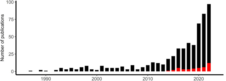
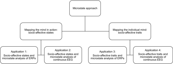
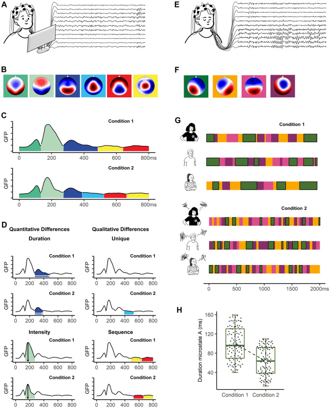
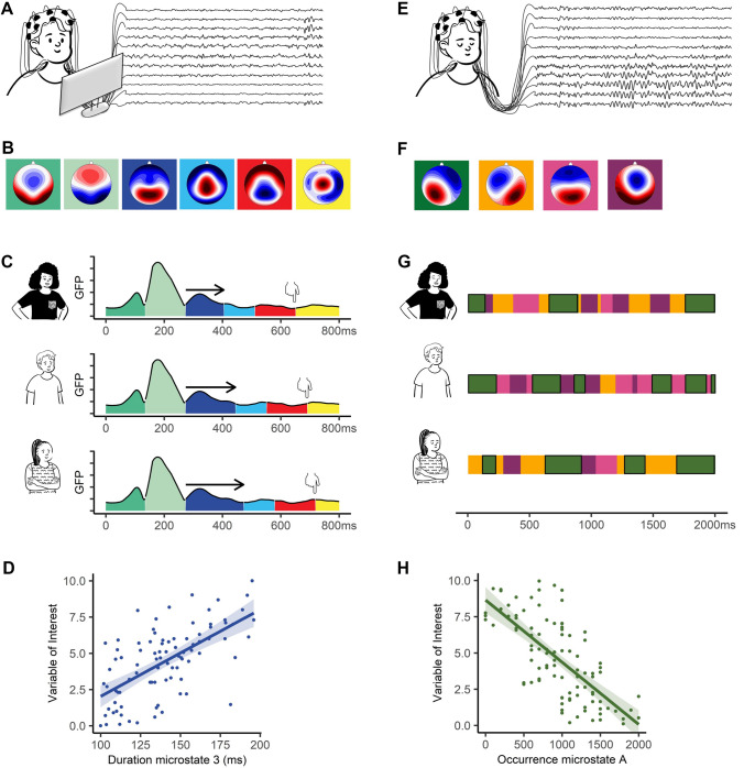
Social interactions require both the rapid processing of multifaceted socio-affective signals (e.g., eye gaze, facial expressions, gestures) and their integration with evaluations, social knowledge, and expectations. Researchers interested in understanding complex social cognition and behavior face a "black box" problem: What are the underlying mental processes rapidly occurring between perception and action and why are there such vast individual differences? In this review, we promote electroencephalography (EEG) microstates as a powerful tool for both examining socio-affective states (e.g., processing whether someone is in need in a given situation) and identifying the sources of heterogeneity in socio-affective traits (e.g., general willingness to help others). EEG microstates are identified by analyzing scalp field maps (i.e., the distribution of the electrical field on the scalp) over time. This data-driven, reference-independent approach allows for identifying, timing, sequencing, and quantifying the activation of large-scale brain networks relevant to our socio-affective mind. In light of these benefits, EEG microstates should become an indispensable part of the methodological toolkit of laboratories working in the field of social and affective neuroscience.

Keywords: Affective neuroscience; EEG microstates; ERP microstates; Neural networks; Social interaction; Social neuroscience.


Conflict of interest statement

The authors declare that they have no conflict of interest.

Figures

Fig. 1
Fig. 1
Number of microstate publications per year. The y-axis displays the number of publications as indexed in PubMed from 1987 to 2022 including the terms “EEG OR ERP AND microstates” (black bars) or the terms “(EEG OR ERP) AND (microstates AND (social OR affective))” (red bars) in the title, keywords, or abstract. Introduced by Dietrich Lehmann in 1987 (Lehmann et al. 1987), microstates initially drew limited attention within the EEG community as indexed by 126 publications over the next 25 years (1987–2012). In the last decade (2013–2022), more than  400 publications have followed. Note that only a minority (around 1/8) of all publications focused on applying microstates to ERP data
Fig. 2
Fig. 2
Overview of the four main applications of EEG microstates. As the first application, one can “map the mind in action” by analyzing ERP microstates evoked by particular socio-affective information. Consider the bus example described at the beginning of this review. ERP microstate analysis opens the “black box” by identifying, timing, and sequencing mental processes occurring between hearing the request by the elderly woman and making the decision to help or not (e.g., neediness evaluation, conflict monitoring, planning behavior). One can then compare these processes’ characteristics across different socio-affective states, for instance, a non-stressed state vs. a stressed state. The second application is to reveal the “socio-affective mind in action” by comparing neural network dynamics extracted from continuous EEG data across a variety of socio-affective states, for instance, during discrete emotions, or during social exclusion vs. social inclusion. The third and fourth application of EEG microstates can be subsumed under the “neural trait approach,” i.e., “mapping the individual mind” by examining socio-affective traits (e.g., Schiller et al., ; for a review, see Nash et al. 2014). Briefly, this approach indexes objective information from stable brain-based characteristics to reveal the sources of individual differences in socio-affective traits (e.g., behavioral intergroup bias, emotion detection ability, prosociality, tendency to deceive others, theory of mind). Microstate parameters from both ERP data (third application) and continuous EEG data (fourth application) are promising “neural trait candidates” as they possess high retest-reliabilities (for ERP: Jouen et al. ; for continuous EEG: Khanna et al. ; Liu et al. ; Schiller et al. ; Antonova et al. ; Kleinert et al. 2023) and heritability (continuous EEG: da Cruz et al. 2020)
Fig. 3
Fig. 3
Mapping the mind in action. Examples of the application of the microstate approach in ERPs (application 1) and in continuous EEG (application 2) are shown on the left and right side, respectively. A ERPs are recorded while participants are confronted with specific socio-affective information (e.g., non-threatening faces vs. threatening faces, non-social vs. social stimuli). B Scalp topographies of 6 clusters in the sequence of their occurrence. C Microstates across time for two conditions plotted over Global Field Power (GFP). D Exemplary quantitative and qualitative differences between the two conditions. E Continuous EEG is recorded while participants are resting with their eyes closed. F Scalp topographies of the 4 prototypical microstate classes during continuous EEG: class A in green, class B in orange, class C in pink, and class D in violet. G Exemplary 2-s of microstates are shown for three individuals in two conditions (e.g., no stress vs. stress; happy mood vs. sad mood). All microstates belonging to class A are highlighted with a black frame. H Participants’ mean durations of microstate class A are shown as box plots for both conditions. The white diamond shape indicates the mean duration in the two conditions, the horizontal line the median. All figure panels are based on simulated data
Fig. 4
Fig. 4
Mapping the individual mind. Examples of the application of the microstate approach in ERPs (application 3) and in continuous EEG (application 4) are shown on the left and right side, respectively. A ERPs are recorded while participants are viewing and responding to some specific socio-affective information (e.g., threatening faces, social stimuli). B Scalp topographies of the 6 clusters in the sequence of their occurrence. C Exemplary individual differences in microstates and response times across time plotted over Global Field Power (GFP). The hand symbols indicate mean response times. Arrows indicate the different durations of the third microstate. D Scatterplot of the association between duration of microstate 3 in milliseconds and the variable of interest (e.g., mean response time, indicated by the hands, in a particular condition of interest). E Continuous EEG is recorded while participants are resting with their eyes closed. F Scalp topographies of the four prototypical microstate classes during resting EEG: class A in green, class B in orange, class C in pink, and class D in violet. G Exemplary 2-s of microstates are shown for three individuals during resting condition. In panel G, all microstates belonging to class A are highlighted with a black frame. H Scatterplot of the association between the occurrence of microstate class A and the variable of interest (e.g., prosocial preferences, behavioral intergroup bias). All figure panels are based on simulated data

References

    1. Antonova E, Holding M, Suen HC, Sumich A, Maex R, Nehaniv C. EEG microstates: functional significance and short-term test-retest reliability. Neuroimage Rep. 2022;2:100089. doi: 10.1016/j.ynirp.2022.100089. - DOI
    1. Asai T, Hamamoto T, Kashihara S, Imamizu H. Real-time detection and feedback of canonical electroencephalogram microstates: validating a neurofeedback system as a function of delay. Front Syst Neurosci. 2022;16:786200. doi: 10.3389/fnsys.2022.786200. - DOI - PMC - PubMed
    1. Bacigalupo F, Luck SJ. Event-related potential components as measures of aversive conditioning in humans. Psychophysiology. 2018;55:e13015. doi: 10.1111/psyp.13015. - DOI - PMC - PubMed
    1. Brandeis D, Lehmann D, Michel CM, Mingrone W. Mapping event-related brain potential microstates to sentence endings. Brain Topogr. 1995;8:145–159. doi: 10.1007/BF01199778. - DOI - PubMed
    1. Bréchet L, Brunet D, Perogamvros L, Tononi G, Michel CM. EEG microstates of dreams. Sci Rep. 2020;10:17069. doi: 10.1038/s41598-020-74075-z. - DOI - PMC - PubMed