

Granuloma annulare on ultrasound: a diagnosis to consider in pediatric skin lesions
- PMID: 37523040
- PMCID: PMC10632325
- DOI: 10.1007/s40477-023-00806-3
Granuloma annulare on ultrasound: a diagnosis to consider in pediatric skin lesions
Abstract
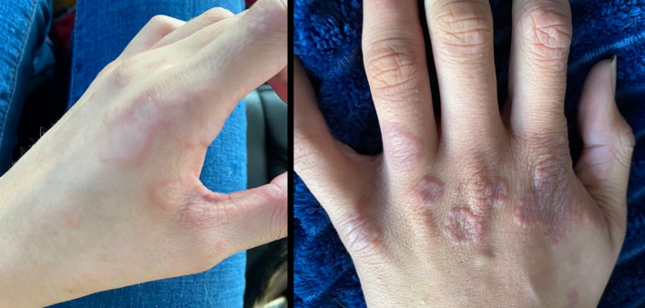
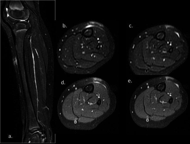
Background: Granuloma annulare (GA) is a rare, benign, inflammatory, self-limited, granulomatous dermatosis that affects children and young adults. The most frequent clinical form is localized GA. Deep GA generally presents as painless palpable subcutaneous nodules in the lower extremities, buttocks, hands and scalp. They may have a fast-growing firm subcutaneous mass presentation, mimicking a malignant lesion which requires an imaging evaluation. Diagnosis of deep GA can be more difficult and imaging evaluation is frequently performed, ultrasound being one of the techniques used.
Objective: To describe the US characteristics of GA in a pediatric series.
Materials and method: Descriptive, retrospective, 14-year study of all pediatrics GA cases.
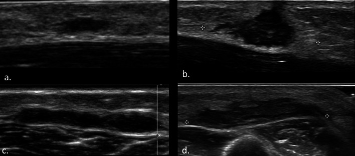
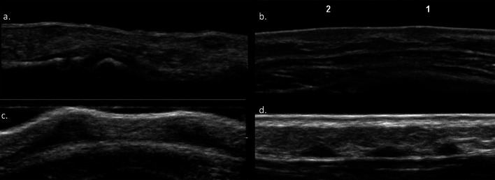
Results: Twelve pediatric cases with GA. 66% females. The lesions were mainly distributed in the extremities: 50% in the lower extremities and 42% in the upper extremities, mostly with multiple lesions. A total of 45 lesions were analyzed, 8 superficial lesions and 37 deep lesions. On ultrasound, the superficial GA corresponded to hypoechoic poorly defined solid plaque like or nodular lesions, located in the dermal-epidermal plane. The deep GA presented as solid nodular, poorly defined hypoechoic lesions that compromised the deep subcutaneous-aponeurotic plane.
Conclusion: GA is an inflammatory lesion that presents as a superficial or deep palpable nodule that predominantly affects children. Superficial and deep GA present characteristic findings on US that can guide the diagnosis. The radiologist needs to know its US appearance to be able to suggest the diagnosis, especially in multiples lesions.
Keywords: Granuloma annulare; Skin lesions; Soft tissue nodules; Ultrasound.
© 2023. Società Italiana di Ultrasonologia in Medicina e Biologia (SIUMB).
Conflict of interest statement
The authors have no relevant financial or non-financial interests to disclose.
Figures


References
-
- Corigliano M, Achenbach RE (2012) Granuloma annulare: a diagnostic and therapeutic challenge. Argentine J Dermatol 93(4). http://www.scielo.org.ar/scielo.php?script=sci_arttext&pid=S1851-300X201...
MeSH terms
LinkOut - more resources
Full Text Sources
