

Lack of orientation specific adaptation to vertically oriented Glass patterns in human visual cortex: an fMRI adaptation investigation
- PMID: 37524748
- PMCID: PMC10390522
- DOI: 10.1038/s41598-023-39247-7
Lack of orientation specific adaptation to vertically oriented Glass patterns in human visual cortex: an fMRI adaptation investigation
Abstract
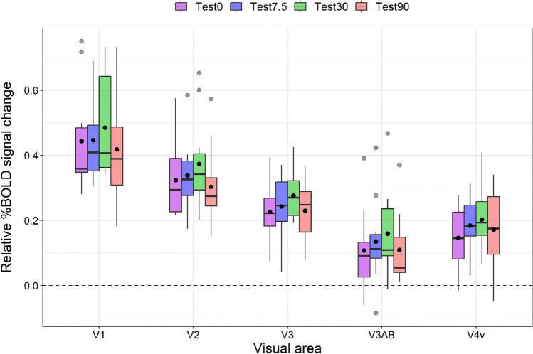
The perception of coherent form configurations in natural scenes relies on the activity of early visual areas that respond to local orientation cues. Subsequently, high-level visual areas pool these local signals to construct a global representation of the initial visual input. However, it is still debated whether neurons in the early visual cortex respond also to global form features. Glass patterns (GPs) are visual stimuli employed to investigate local and global form processing and consist of randomly distributed dots pairs called dipoles arranged to form specific global configurations. In the current study, we used GPs and functional magnetic resonance imaging (fMRI) adaptation to reveal the visual areas that subserve the processing of oriented GPs. Specifically, we adapted participants to vertically oriented GP, then we presented test GPs having either the same or different orientations with respect to the adapting GP. We hypothesized that if local form features are processed exclusively by early visual areas and global form by higher-order visual areas, then the effect of visual adaptation should be more pronounced in higher tier visual areas as it requires global processing of the pattern. Contrary to this expectation, our results revealed that adaptation to GPs is robust in early visual areas (V1, V2, and V3), but not in higher tier visual areas (V3AB and V4v), suggesting that form cues in oriented GPs are primarily derived from local-processing mechanisms that originate in V1. Finally, adaptation to vertically oriented GPs causes a modification in the BOLD response within early visual areas, regardless of the relative orientations of the adapting and test stimuli, indicating a lack of orientation selectivity.
© 2023. The Author(s).
Conflict of interest statement
The authors declare no competing interests.
Figures


References
Publication types
MeSH terms
LinkOut - more resources
Full Text Sources
Medical
