

Evaluation of a 3D-printed hands-on radius fracture model during teaching courses
- PMID: 37524864
- PMCID: PMC10923998
- DOI: 10.1007/s00068-023-02327-4
Evaluation of a 3D-printed hands-on radius fracture model during teaching courses
Abstract
Objective: This study aimed to evaluate the effectiveness of a 3D-printed hands-on radius fracture model for teaching courses. The model was designed to enhance understanding and knowledge of radius fractures among medical students during their clinical training.
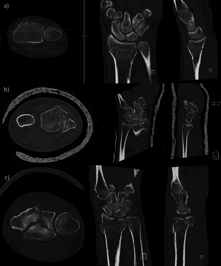
Methods: The 3D models of radius fractures were generated using CT scans and computer-aided design software. The models were then 3D printed using Fused-Filament-Fabrication (FFF) technology. A total of 170 undergraduate medical students participated in the study and were divided into three groups. Each group was assigned one of three learning aids: conventional X-ray, CT data, or a 3D-printed model. After learning about the fractures, students completed a questionnaire to assess their understanding of fracture mechanisms, ability to assign fractures to the AO classification, knowledge of surgical procedures, and perception of the teaching method as well as the influence of such courses on their interest in the specialty of trauma surgery. Additionally, students were tested on their ability to allocate postoperative X-ray images to the correct preoperative image or model and to classify them to the AO classification.
Results: The 3D models were well received by the students, who rated them as at least equal or better than traditional methods such as X-ray and CT scans. Students felt that the 3D models improved their understanding of fracture mechanisms and their ability to explain surgical procedures. The results of the allocation test showed that the combination of the 3D model and X-ray yielded the highest accuracy in classifying fractures according to the AO classification system, although the results were not statistically significant.
Conclusion: The 3D-printed hands-on radius fracture model proved to be an effective teaching tool for enhancing students' understanding of fracture anatomy. The combination of 3D models with the traditional imaging methods improved students' ability to classify fractures and allocate postoperative images correctly.
Keywords: 3D printing; Fracture models; Radius fractures; Teaching; Training; Traumatology.
© 2023. The Author(s).
Conflict of interest statement
The authors declare that they have no conflict of interest.
Figures


References
-
- Neijhoft J, Viertmann T, Meier S, Söhling N, Wicker S, Henrich D, Marzi I. Manufacturing and supply of face shields in hospital operation in case of unclear and confirmed COVID-19 infection status of patients. Eur J Trauma Emerg Surg. 2020;46:743–745. doi: 10.1007/s00068-020-01392-3. - DOI - PMC - PubMed
-
- Rundgren J, Bojan A, Mellstrand Navarro C, Enocson A. Epidemiology, classification, treatment and mortality of distal radius fractures in adults: an observational study of 23,394 fractures from the national Swedish fracture register. BMC Musculoskelet Disord. 2020;21:88. doi: 10.1186/s12891-020-3097-8. - DOI - PMC - PubMed
MeSH terms
LinkOut - more resources
Full Text Sources
Medical
