. 2023 Aug;55(8):1743-1756.
doi: 10.1038/s12276-023-01059-0. Epub 2023 Aug 1.

ALKBH5 facilitates CYP1B1 mRNA degradation via m6A demethylation to alleviate MSC senescence and osteoarthritis progression

Affiliations

ALKBH5 facilitates CYP1B1 mRNA degradation via m6A demethylation to alleviate MSC senescence and osteoarthritis progression

Guiwen Ye et al. Exp Mol Med. 2023 Aug.

Abstract

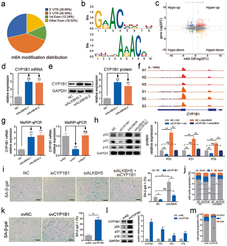
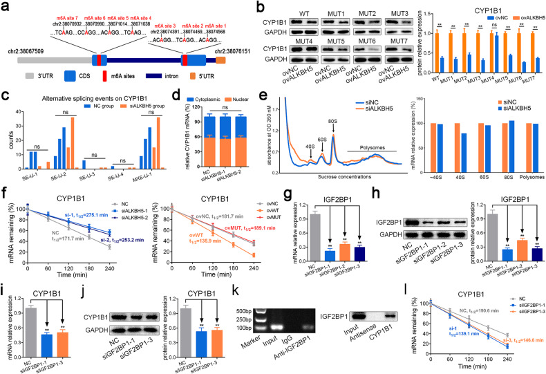
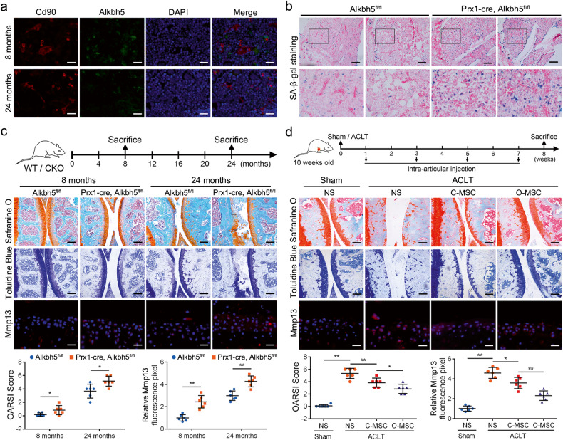
Improving health and delaying aging is the focus of medical research. Previous studies have shown that mesenchymal stem cell (MSC) senescence is closely related to organic aging and the development of aging-related diseases such as osteoarthritis (OA). m6A is a common RNA modification that plays an important role in regulating cell biological functions, and ALKBH5 is one of the key m6A demethylases. However, the role of m6A and ALKBH5 in MSC senescence is still unclear. Here, we found that the m6A level was enhanced and ALKBH5 expression was decreased in aging MSCs induced by multiple replications, H2O2 stimulation or UV irradiation. Downregulation of ALKBH5 expression facilitated MSC senescence by enhancing the stability of CYP1B1 mRNA and inducing mitochondrial dysfunction. In addition, IGF2BP1 was identified as the m6A reader restraining the degradation of m6A-modified CYP1B1 mRNA. Furthermore, Alkbh5 knockout in MSCs aggravated spontaneous OA in mice, and overexpression of Alkbh5 improved the efficacy of MSCs in OA. Overall, this study revealed a novel mechanism of m6A in MSC senescence and identified promising targets to protect against aging and OA.


Conflict of interest statement

The authors declare no competing interests.

Figures

Fig. 1
Fig. 1. The level of m6A modification increased, and METTL3 and ALKBH5 levels were reduced during MSC senescence.
a The percentages of SA-β-gal-positive cells increased with cell replication. b The protein levels of p53, p21 and p16 increased with cell replication. c The level of m6A modification increased with cell replication. d H2O2 treatment enhanced the percentages of SA-β-gal-positive cells. e H2O2 treatment enhanced the protein levels of p53, p21 and p16. f H2O2 treatment enhanced the level of m6A modification. g UV irradiation enhanced the percentages of SA-β-gal-positive cells. h UV irradiation enhanced the protein levels of p53, p21 and p16. i UV irradiation enhanced the level of m6A modification. j The mRNA levels of METTL3 and ALKBH5 decreased with cell replication. k The protein levels of METTL3 and ALKBH5 decreased with cell replication. n = 9, * indicates P < 0.05, ** indicates P < 0.01, ns indicates not significant, scale bar = 50 nm.
Fig. 2
Fig. 2. ALKBH5 deficiency facilitated MSC senescence.
a The silencing efficiency of siRNAs targeting METTL3. b The silencing efficiency of siRNAs targeting ALKBH5. c, d Knockdown of ALKBH5 increased the percentages of SA-β-gal-positive cells, but knockdown of METTL3 had no significant effect (scale bar = 50 nm). e Knockdown of ALKBH5 increased the protein levels of p53, p21 and p16, and knockdown of METTL3 decreased the protein levels of p21 but had no significant effect on the protein levels of p53 or p16. f Knockdown of ALKBH5 increased the percentages of G0/G1 cells in the cell cycle, but knockdown of METTL3 had no significant effect. g Knockdown of ALKBH5 enhanced the p-H2A.X signal, but knockdown of METTL3 had no significant effect. Scale bar = 20 nm. n = 9, * indicates P < 0.05, ** indicates P < 0.01, ns indicates not significant.
Fig. 3
Fig. 3. ALKBH5 alleviated MSC senescence via m6A demethylation activity.
a The overexpression efficiency of WT-ALKBH5 and MUT-ALKBH5. b WT-ALKBH5 reduced the percentages of SA-β-gal-positive cells, but MUT-ALKBH5 had no significant effect. c WT-ALKBH5 reduced the protein levels of p53, p21 and p16, but MUT-ALKBH5 had no significant effect. d WT-ALKBH5 reduced the percentages of G0/G1 in the cell cycle, but MUT-ALKBH5 had no significant effect. e WT-ALKBH5 improved the effect of H2O2 on SA-β-gal staining, but MUT-ALKBH5 had no significant effect. f WT-ALKBH5 improved the effect of H2O2 on the protein levels of p53, p21 and p16, but MUT-ALKBH5 had no significant effect. g WT-ALKBH5 improved the effect of H2O2 on the percentages of G0/G1 in the cell cycle, but MUT-ALKBH5 had no significant effect. h WT-ALKBH5 improved the effect of UV on SA-β-gal staining, but MUT-ALKBH5 had no significant effect. i WT-ALKBH5 improved the effect of UV on the protein levels of p53, p21 and p16, but MUT-ALKBH5 had a nonsignificant effect. j WT-ALKBH5 improved the effect of UV on the percentages of G0/G1 in the cell cycle, but MUT-ALKBH5 had no significant effect. n = 9, * indicates P < 0.05, ** indicates P < 0.01, ns indicates not significant, scale bar = 50 nm.
Fig. 4
Fig. 4. CYP1B1 is the downstream target of ALKBH5 in senescence regulation.
a The m6A modification distribution of the MeRIP-Seq data. b The consensus sequence motifs for m6A methylation in the MeRIP-Seq. c The conjoint analysis of m6A modification and gene expression. d Knockdown of ALKBH5 increased the mRNA level of CYP1B1. e Knockdown of ALKBH5 increased the protein level of CYP1B1. f The m6A peaks diagram showed that the m6A abundance on CYP1B1 mRNA increased in the ALKBH5-silencing group. g MeRIP-qPCR showed that knockdown of ALKBH5 increased and overexpression of WT-ALKBH5 reduced the level of m6A modification on CYP1B1 mRNA but MUT-ALKBH5 had no significant effect. h Knockdown of CYP1B1 reduced the protein levels of p53, p21 and p16 and rescued the effect of ALKBH5 knockdown on them. i Knockdown of CYP1B1 reduced the percentages of SA-β-gal-positive cells and rescued the effect of ALKBH5 knockdown. j Knockdown of CYP1B1 reduced the percentages of G0/G1 cells in the cell cycle and rescued the effect of ALKBH5 knockdown. k Overexpression of CYP1B1 increased the percentage of SA-β-gal-positive cells. l Overexpression of CYP1B1 increased the protein levels of p53, p21 and p16. m Overexpression of CYP1B1 increased the percentage of G0/G1 cells in the cell cycle. n = 9, * indicates P < 0.05, ** indicates P < 0.01, ns indicates not significant, scale bar = 50 nm.
Fig. 5
Fig. 5. ALKBH5 regulated MSC senescence though CYP1B1 via mitochondrial dysfunction.
a Overexpression of CYP1B1 increased the level of ROS. b Knockdown of CYP1B1 reduced the level of ROS and rescued the effect of ALKBH5 knockdown. c, d Overexpression of CYP1B1 impaired the MMP, and knockdown of CYP1B1 elevated the MMP and rescued the effect of ALKBH5 knockdown, scale bar = 25 nm. e Overexpression of CYP1B1 impaired the level of ATP, and knockdown of CYP1B1 elevated the level of ATP and rescued the effect of ALKBH5 knockdown. f Overexpression of CYP1B1 impaired the signal of MitoSOX, scale bar = 50 nm. g Knockdown of CYP1B1 elevated the signal of MitoSOX and rescued the effect of ALKBH5 knockdown. Scale bar = 50 nm. n = 9, * indicates P < 0.05, ** indicates P < 0.01, ns indicates not significant.
Fig. 6
Fig. 6. Knockdown of ALKBH5 enhanced CYP1B1 mRNA stability in an IGF2BP1-dependent manner.
a The m6A sites predicted with high confidence on CYP1B1 mRNA. b Overexpression of ALKBH5 reduced the protein levels of CYP1B1 in the WT-CYP1B1 group and other MUT groups except for the MUT4 group. c The alternative splicing events of CYP1B1 analyzed from the RNA-Seq data by rMATS. d The nuclear and cytoplasmic distribution of CYP1B1 mRNA was not changed significantly with knockdown of ALKBH5. e The abundance of CYP1B1 mRNA on polysomes was not changed significantly with knockdown of ALKBH5. f Knockdown of ALKBH5 reduced the degradation rate of CYP1B1 mRNA, and WT-ALKBH5 facilitated the degradation rate of CYP1B1 mRNA but MUT-ALKBH5 had no significant effect. g The silencing efficiency of siRNAs targeting IGF2BP1 at the mRNA level. h The silencing efficiency of siRNAs targeting IGF2BP1 at the protein level. i Knockdown of IGF2BP1 reduced the mRNA level of CYP1B1. j Knockdown of IGF2BP1 reduced the protein level of CYP1B1. k RIP assays showed that CYP1B1 mRNA was precipitated by IGF2BP1 antibody (left), and RNA pulldown assays showed that IGF2BP1 protein was pulled down by CYP1B1 mRNA (right). l Knockdown of IGF2BP1 facilitated the degradation of CYP1B1 mRNA. n = 9, ** indicates P < 0.01, ns indicates not significant.
Fig. 7
Fig. 7. Conditional Alkbh5 knockout mice displayed a severe OA phenotype, and Alkbh5 improved the efficacy of MSCs in ACLT-induced OA.
a The Alkbh5 signal on Cd105-positive cells was impaired in 24-month-old mice compared to 8-month-old mice; scale bar = 40 nm. b SA-β-gal staining was stronger in the Prx1-cre; Alkbh5fl/fl mice than in the Alkbh5fl/fl control littermates; scale bar = 200 nm. c Both at 8 months and 24 months, the OARSI score and Mmp13 signal of the knee joint were increased in the Prx1-cre; Alkbh5fl/fl mice compared to the Alkbh5fl/fl mice. d The knee joints of the ACLT group mice had more severe OARSI scores and higher Mmp13 expression. Intra-articular injection of control MSCs (C-MSCs) improved the OARSI score and Mmp13 expression, while overexpressing ALKBH5 MSCs (O-MSCs) had a better effect; scale bar = 200 nm (top and middle), scale bar = 40 nm (bottom). n = 6, * indicates P < 0.05, ** indicates P < 0.01, ns indicates not significant.

References

    1. He S, Sharpless NE. Senescence in health and disease. Cell. 2017;169:1000–1011. doi: 10.1016/j.cell.2017.05.015. - DOI - PMC - PubMed
    1. Muñoz-Espín D, Serrano M. Cellular senescence: from physiology to pathology. Nat. Rev. Mol. Cell Biol. 2014;15:482–96. doi: 10.1038/nrm3823. - DOI - PubMed
    1. Hernandez-Segura A, Nehme J, Demaria M. Hallmarks of cellular senescence. Trends Cell Biol. 2018;28:436–453. doi: 10.1016/j.tcb.2018.02.001. - DOI - PubMed
    1. Wang W, et al. A genome-wide CRISPR-based screen identifies KAT7 as a driver of cellular senescence. Sci. Transl. Med. 2021;13:eabd2655. doi: 10.1126/scitranslmed.abd2655. - DOI - PubMed
    1. Ding DC, Shyu WC, Lin SZ. Mesenchymal stem cells. Cell Transplant. 2011;20:5–14. doi: 10.3727/096368910X. - DOI - PubMed

Publication types