. 2023 Jul 31;10(1):35.
doi: 10.1186/s40779-023-00469-5.

The marriage of immunomodulatory, angiogenic, and osteogenic capabilities in a piezoelectric hydrogel tissue engineering scaffold for military medicine

Affiliations

The marriage of immunomodulatory, angiogenic, and osteogenic capabilities in a piezoelectric hydrogel tissue engineering scaffold for military medicine

Ping Wu et al. Mil Med Res. .

Abstract

Background: Most bone-related injuries to grassroots troops are caused by training or accidental injuries. To establish preventive measures to reduce all kinds of trauma and improve the combat effectiveness of grassroots troops, it is imperative to develop new strategies and scaffolds to promote bone regeneration.

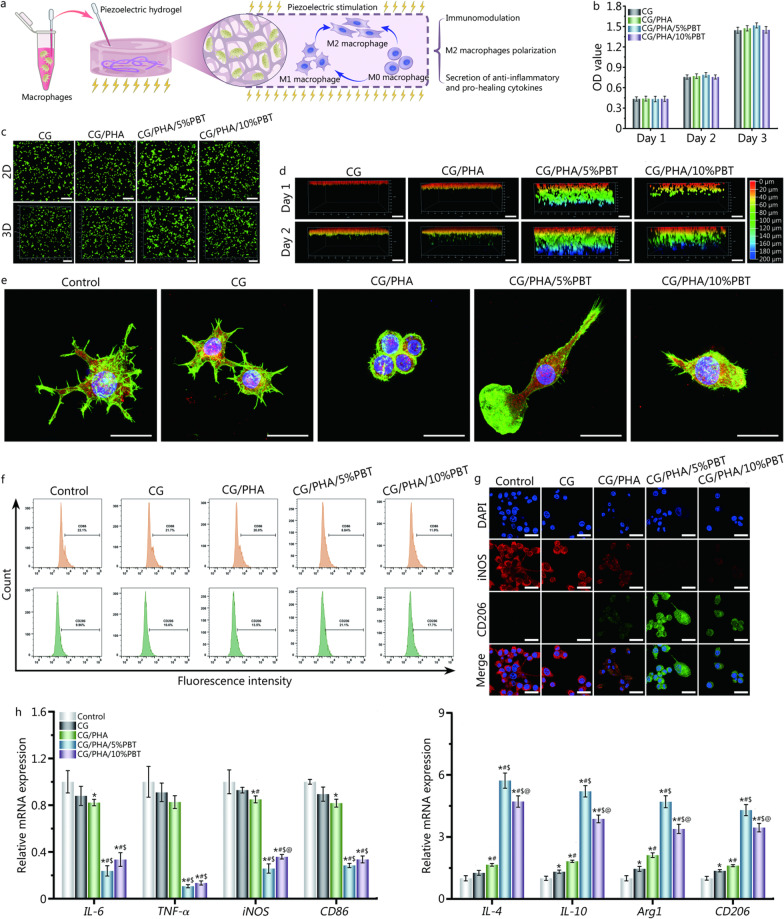
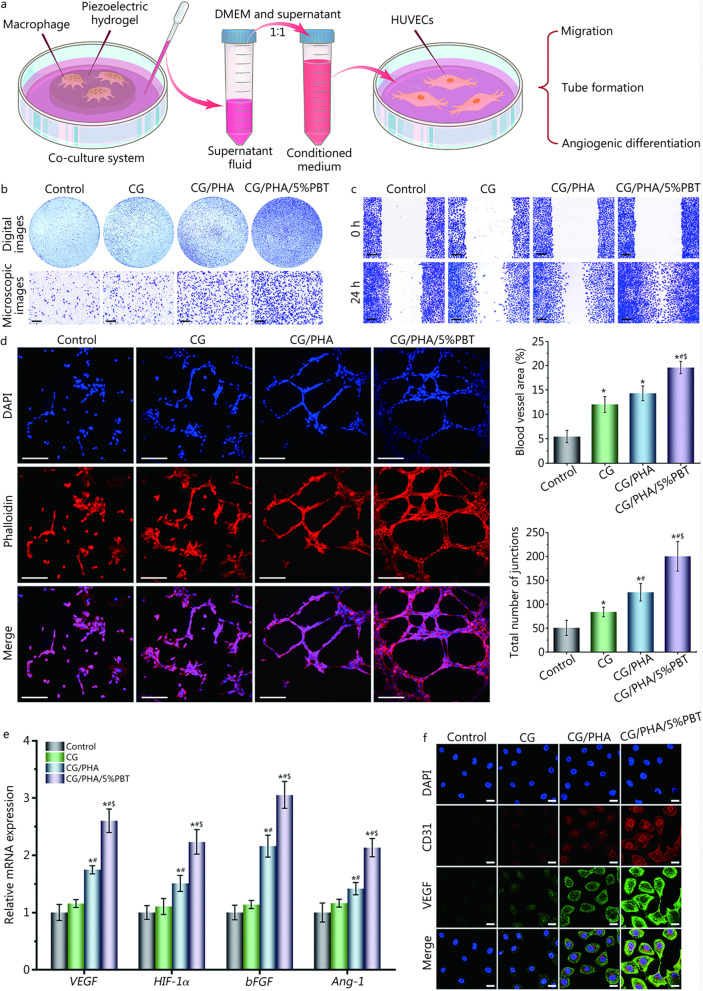
Methods: In this study, a porous piezoelectric hydrogel bone scaffold was fabricated by incorporating polydopamine (PDA)-modified ceramic hydroxyapatite (PDA-hydroxyapatite, PHA) and PDA-modified barium titanate (PDA-BaTiO3, PBT) nanoparticles into a chitosan/gelatin (Cs/Gel) matrix. The physical and chemical properties of the Cs/Gel/PHA scaffold with 0-10 wt% PBT were analyzed. Cell and animal experiments were performed to characterize the immunomodulatory, angiogenic, and osteogenic capabilities of the piezoelectric hydrogel scaffold in vitro and in vivo.

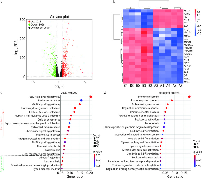
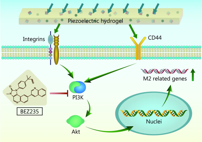
Results: The incorporation of BaTiO3 into the scaffold improved its mechanical properties and increased self-generated electricity. Due to their endogenous piezoelectric stimulation and bioactive constituents, the as-prepared Cs/Gel/PHA/PBT hydrogels exhibited cytocompatibility as well as immunomodulatory, angiogenic, and osteogenic capabilities; they not only effectively induced macrophage polarization to M2 phenotype but also promoted the migration, tube formation, and angiogenic differentiation of human umbilical vein endothelial cells (HUVECs) and facilitated the migration, osteo-differentiation, and extracellular matrix (ECM) mineralization of MC3T3-E1 cells. The in vivo evaluations showed that these piezoelectric hydrogels with versatile capabilities significantly facilitated new bone formation in a rat large-sized cranial injury model. The underlying molecular mechanism can be partly attributed to the immunomodulation of the Cs/Gel/PHA/PBT hydrogels as shown via transcriptome sequencing analysis, and the PI3K/Akt signaling axis plays an important role in regulating macrophage M2 polarization.

Conclusion: The piezoelectric Cs/Gel/PHA/PBT hydrogels developed here with favorable immunomodulation, angiogenesis, and osteogenesis functions may be used as a substitute in periosteum injuries, thereby offering the novel strategy of applying piezoelectric stimulation in bone tissue engineering for the enhancement of combat effectiveness in grassroots troops.

Keywords: Angiogenesis; Immunomodulation; Osteogenic differentiation; Piezoelectric hydrogel; Tissue engineering scaffold.


Conflict of interest statement

The authors declare that they have no known competing financial interests or personal relationships that could influence the work reported in this paper.

Figures

Fig. 1
Fig. 1
Schematic illustration of the design strategy of the piezoelectric CG/PHA/PBT hydrogels and the bone regeneration mechanism. a The preparation process of the piezoelectric CG/PHA/PBT hydrogels. b The application and potential biological mechanism of the piezoelectric hydrogels for rapid bone regeneration. c Schematic illustration of the possible self-powered mechanism of the piezoelectric CG/PHA/PBT hydrogels. The dipoles in the BT nanoparticles are oriented in the same direction in the piezoelectric hydrogels. The electric polarization is presented in the direction of the oriented dipoles and can produce a piezoelectric potential in the BT piezoelectric nanoparticles. In the absence of external stimuli, there is a positive and negative charge balance in the hydrogel. Once a pressure stimulus is applied to the piezoelectric hydrogels, the electrons in the hydrogels flow out of the hydrogel. Once the pressure disappears, the cumulative free charges flow back into the piezoelectric hydrogels. BT barium titanate, HA hydroxyapatite, Cs chitosan, Gel gelatin, PHA polydopamine coated-hydroxyapatite, PBT polydopamine coated-barium titanate, e electron, R resistance, M0 mφ unpolarized macrophage, M1 mφ type 1 macrophage, M2 mφ type 2 macrophage, IL-6 interleukin-6, IL-4 interleukin-4, TNF-α tumor necrosis factor-α, iNOS inducible nitric-oxide synthase, CD86 cluster of differentiation 86 protein, Arg1 arginase, BMSC bone marrow stromal cell, HUVEC human umbilical vein endothelial cell
Fig. 2
Fig. 2
Characterization of the piezoelectric hydrogels. a Representative SEM images of different hydrogel samples, the red arrows represent piezoelectric nanoparticles. b EDS elemental mapping of the CG/PHA/5%PBT piezoelectric hydrogel. XRD (c), FTIR (d), rheological curve (e), and elasticity modulus (f) of different hydrogel samples. g The output voltage of different hydrogel samples under 10 Hz and 1 kP pressure. *P < 0.05, compared with the CG group; #P < 0.05, compared with the CG/PHA group. CG chitosan/gelatin, PHA polydopamine coated-hydroxyapatite, PBT polydopamine coated-barium titanate, SEM scanning electron microscope, EDS energy disperse spectroscopy, XRD X-ray diffraction, FTIR fourier transform infrared spectroscopy
Fig. 3
Fig. 3
In vitro evaluation of the immunomodulatory properties of the piezoelectric hydrogels. a Schematic diagram of immune regulation by the piezoelectric hydrogels. b Evaluation of the cytocompatibility of the piezoelectric hydrogels. c–d 2D and 3D live and dead cell staining of macrophages (RAW 264.7 cells) cocultured with the piezoelectric hydrogels. Scale bar: 200 μm (c), 100 μm (d). e Representative immunofluorescence images of RAW 264.7 cells on hydrogels. F-actin (green) is a fibrous actin that presents the skeleton of the cells, F4/80 (red) is cell surface glycoprotein and the marker of mature mouse macrophages, DAPI (blue) stands for the nucleus. Scale bar: 25 μm. f Representative flow cytometric dot plots showing cell surface markers of RAW 264.7 cells, including CD86 and CD206. g Representative immunofluorescence images of iNOS (red) and CD206 (green) in RAW 264.7 cells on hydrogels on day 2. Scale bar: 25 μm. h Relative mRNA expression levels of anti-inflammatory genes and pro-inflammatory genes of macrophage under the piezoelectric hydrogels stimulation for 2 d. *P < 0.05, compared with the control group; #P < 0.05, compared with the CG group; $P < 0.05, compared with the CG/PHA group; @P < 0.05, compared with the CG/PHA/5%PBT group. CG chitosan/gelatin, PHA polydopamine coated-hydroxyapatite, PBT polydopamine coated-barium titanate, IL-6 interleukin-6, TNF-α tumor necrosis factor-α, iNOS inducible nitric-oxide synthase, IL-4 interleukin-4, IL-10 interleukin-10, Arg1 arginase 1
Fig. 4
Fig. 4
In vitro assessment of the angiogenic ability of the piezoelectric hydrogels. a Schematic diagram of the pro-angiogenic cell experiment. First, the piezoelectric hydrogel was co-cultured with macrophages; then, vascular endothelial cells were cultured using the co-culture medium from macrophages. b Representative digital and microscopic images of the migrated HUVECs cells to the lower chamber after the macrophage medium was separately added to the lower chamber and cultured for 24 h. Scale bar: 200 μm. c Representative microscopic images of HUVECs after the cells were co-cultured with different macrophage media for 24 h. Scale bar: 200 μm. d Representative fluorescence images and quantitative analysis of HUVEC tube formation after the cells were co-cultured with different macrophage media for 8 h. Percentage of blood vessel area is the vessel density of vascular endothelial cells in a single image, total number of junctions is the cell intersections between vascular endothelial cells in a single image, tube formation was quantified using AngioTool (National Cancer Institute, NIH) for the percentage of blood vessel area and the total number of junctions. Scale bar: 250 μm. e Relative mRNA expression levels of angiogenesis-related genes in HUVECs after the cells were co-cultured with different macrophage media for 7 d, including VEGF, HIF-1α, bFGF, and Ang-1. f Representative immunofluorescence images of CD31 (red), VEGF (green), and nuclei (blue) in HUVECs after the cells were co-cultured with different macrophage media for 7 d. Scale bar: 25 μm. *P < 0.05, compared with the control group; #P < 0.05, compared with the CG group; $ P < 0.05, compared with the CG/PHA group. CG chitosan/gelatin, PHA polydopamine coated-hydroxyapatite, PBT polydopamine coated-barium titanate, VEGF vascular endothelial growth factor, HIF-1α hypoxia inducible factor-1α, bFGF basic fibroblast growth factor, Ang-1 angiopoietin-1, HUVECs human umbilical vein endothelial cells
Fig. 5
Fig. 5
The CG/PHA/5%PBT piezoelectric hydrogel promotes bone tissue regeneration in vivo. a 2D micro-CT coronal and sagittal images, and the BV/TV and BMD results of regenerated bone tissues. Scale bar: 200 μm. b HE, Masson’s trichrome, and Goldner’s trichrome staining of the bone defect 8 weeks after implantation. Scale bar: 200 μm. Yellow arrows and black arrows represent the newly formed central canal and bone lacunae within the defect region, respectively. For Goldner’s trichrome staining images, a small island of immature bone (osteoid, red) was detected at the periphery of the defect site in the control group. Black dotted lines define the boundary of the critical-sized cranial bone defect. *P < 0.05, compared with the control group; #P < 0.05, compared with the CG group; $P < 0.05, compared with the CG/PHA group. CG chitosan/gelatin, PHA polydopamine coated-hydroxyapatite, PBT polydopamine coated-barium titanate, BV/TV bone tissue volume/total tissue volume, BMD bone mineral density, FT fibrous tissue, NB newly formed bone tissue, HB host bone, MB mineralized/mature bone
Fig. 6
Fig. 6
Transcriptome analysis of macrophage immunity regulated by the CG/PHA/5%PBT piezoelectric hydrogel. a Quantitative analysis of DEGs in macrophages from the CG and CG/PHA/5%PBT groups. b Heatmap of DEGs associated with immune regulation. A1–A5 are CG hydrogel samples, and B1–B5 are CG/PHA/5%PBT piezoelectric hydrogel samples. c KEGG pathway analysis in macrophages from the CG and CG/PHA/5%PBT groups. d GO enrichment analysis of the 20 most differentially up-regulated and down-regulated biological processes. CG chitosan/gelatin, PHA polydopamine coated-hydroxyapatite, PBT polydopamine coated-barium titanate, DEGs differentially expressed genes, KEGG kyoto encyclopedia of genes and genomes, GO gene ontology
Fig. 7
Fig. 7
Schematic diagram of the possible molecular mechanism by which the CG/PHA/5%PBT piezoelectric hydrogel promotes bone repair by regulating macrophage M2 polarization and activating the PI3K/Akt axis. CG chitosan/gelatin, PHA polydopamine coated-hydroxyapatite, PBT polydopamine coated-barium titanate, PI3K phosphoinositol 3 kinase, Akt serine/threonine protein kinase

References

    1. Xu PC, Xuan M, Cheng B. Effects and mechanism of platelet-rich plasma on military drill injury: a review. Mil Med Res. 2020;7(1):56. - PMC - PubMed
    1. Mathieu L, Mourtialon R, Durand M, de Rousiers A, de L'Escalopier N, Collombet JM. Masquelet technique in military practice: specificities and future directions for combat-related bone defect reconstruction. Mil Med Res. 2022;9(1):48. - PMC - PubMed
    1. Arif ZU, Khalid MY, Noroozi R, Sadeghianmaryan A, Jalalvand M, Hossain M. Recent advances in 3D-printed polylactide and polycaprolactone-based biomaterials for tissue engineering applications. Int J Biol Macromol. 2022;218:930–968. doi: 10.1016/j.ijbiomac.2022.07.140. - DOI - PubMed
    1. Arif ZU, Khalid MY, Ahmed W, Arshad H. A review on four-dimensional (4D) bioprinting in pursuit of advanced tissue engineering applications. Bioprinting. 2022;27:e00203. doi: 10.1016/j.bprint.2022.e00203. - DOI
    1. Zhang J, Tong D, Song H, Ruan R, Sun Y, Lin Y, et al. Osteoimmunity-regulating biomimetically hierarchical scaffold for augmented bone regeneration. Adv Mater. 2022;34(36):e2202044. doi: 10.1002/adma.202202044. - DOI - PubMed

LinkOut - more resources