. 2024 Mar;30(3):e14385.
doi: 10.1111/cns.14385. Epub 2023 Jul 31.

Acoustic-electric trigeminal-nerve stimulation enhances functional connectivity in patients with disorders of consciousness

Affiliations

Acoustic-electric trigeminal-nerve stimulation enhances functional connectivity in patients with disorders of consciousness

Min Wu et al. CNS Neurosci Ther. 2024 Mar.

Abstract

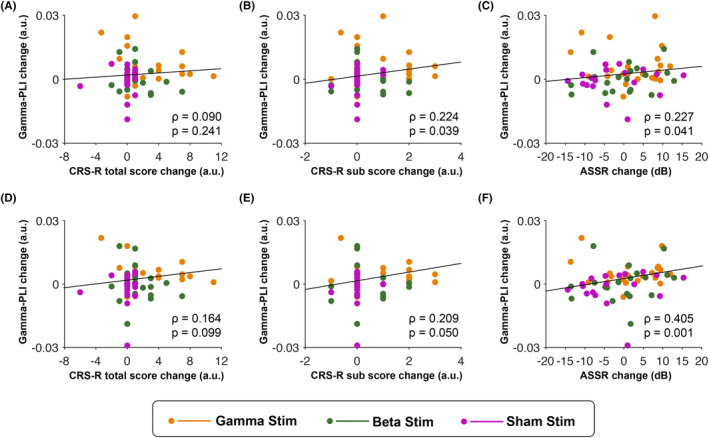
Aim: Disruption of functional brain connectivity is thought to underlie disorders of consciousness (DOC) and recovery of impaired connectivity is suggested as an indicator of consciousness restoration. We recently found that rhythmic acoustic-electric trigeminal-nerve stimulation (i.e., musical stimulation synchronized to electrical stimulation of the trigeminal nerve) in the gamma band can improve consciousness in patients with DOC. Here, we investigated whether these beneficial stimulation effects are mediated by alterations in functional connectivity.

Methods: Sixty-three patients with DOC underwent 5 days of gamma, beta, or sham acoustic-electric trigeminal-nerve stimulation. Resting-state electroencephalography was measured before and after the stimulation and functional connectivity was assessed using phase-lag index (PLI).

Results: We found that gamma stimulation induces an increase in gamma-band PLI. Further characterization revealed that the enhancing effect is (i) specific to the gamma band (as we observed no comparable change in beta-band PLI and no effect of beta-band acoustic-electric stimulation or sham stimulation), (ii) widely spread across the cortex, and (iii) accompanied by improvements in patients' auditory abilities.

Conclusion: These findings show that gamma acoustic-electric trigeminal-nerve stimulation can improve resting-state functional connectivity in the gamma band, which in turn may be linked to auditory abilities and/or consciousness restoration in DOC patients.

Keywords: acoustic stimulation; disorders of consciousness; electric trigeminal-nerve stimulation; gamma band; resting-state functional connectivity.


Conflict of interest statement

All authors declare no competing interests.

Figures

FIGURE 1
FIGURE 1
Schematic of experimental procedure. (A) Each patient underwent a 15‐day long experimental procedure consisting of three 5‐day long consecutive phases: pretest, stimulation, and posttest. The pretest and posttest phases involved administration of daily behavioral assessments (CRS‐R) and a single neural assessment (ca. 55 min EEG) on the day immediately before and after the stimulation phase, respectively. (B) The stimulation phase involved daily administration of 40 min of rhythmic gamma (left) or beta (center) acoustic‐electric trigeminal nerve stimulation, or sham (right) stimulation. CRS‐R, Coma Recovery Scale‐Revised; EEG, electroencephalography.
FIGURE 2
FIGURE 2
Resting‐state functional connectivity in patients with disorders of consciousness (DOC) in the gamma and beta band before and after rhythmic acoustic‐electric or sham trigeminal nerve stimulation. (A) Whole‐scalp gamma phase‐lag index (PLI) in DOC patients in pretest (blue) and posttest (red). The leftmost pair of plots represents the group of patients who underwent gamma stimulation (gamma‐stimulation group). The middle and rightmost pairs represent the patient group receiving beta stimulation (beta‐stimulation group) and sham stimulation (sham‐stimulation group), respectively. Only gamma stimulation was found to have a strengthening effect on patients' brain connectivity in the gamma band, which was significantly larger than the non‐significant change in the beta and in the sham‐stimulation groups. (B). Same as (A), but for brain connectivity in the beta band. Neither acoustic‐electric nor sham stimulation had an effect on patients' brain connectivity in the beta band. The raincloud plots visualize the data distribution, the horizontal line within each boxplot indicates the median value across participants; the bottom and top edges of the box indicate the first and third quartile values; the whiskers indicate the most extreme values within 1.5 times the interquartile range. N.S., non‐significant, *p < 0.05, **p < 0.01.
FIGURE 3
FIGURE 3
Scalp topography of gamma‐band connectivity in patients with disorders of consciousness (DOC) before and after gamma acoustic‐electric trigeminal nerve stimulation. The spatial distribution of gamma‐band connectivity is depicted in lateral (top row), anterior and posterior (middle row), and dorsal and ventral (bottom row) views of the cerebral cortex. The colored nodes represent the locations of the 63 electroencephalographic electrodes on the scalp. Purple, blue, yellow, and cyan nodes, respectively, represent regions of interest (ROIs) over the frontal, central, temporal, and parietooccipital cortex. The red lines represent gamma‐band connections between pairs of nodes. The line thickness represents the strength of the connection (with thicker lines representing higher phase‐lag index (PLI) values, i.e., stronger connection). A threshold PLI value of 0.085 was applied for clear visualization. (A) Before gamma stimulation, DOC patients were observed to show almost no suprathreshold gamma‐band connectivity within or between any ROIs. (B) After receiving the gamma stimulation, patients showed enhanced gamma‐band connectivity within and between widespread ROIs.
FIGURE 4
FIGURE 4
Effect of gamma acoustic‐electric trigeminal nerve stimulation on gamma‐band connectivity in patients with disorders of consciousness (DOC) within and between regions of interest (ROIs). Each cell represents the change in gamma‐band connectivity within a given ROI (cells on the diagonal) or between a given pair of ROIs (cells off the diagonal) after gamma stimulation (pretest vs. posttest). The color of the cell represents the effect size of the change, with darker colors representing stronger phase‐lag index increases. Asterisks represent the statistical significance of the change (N.S., non‐significant, *p < 0.05, **p < 0.01, false discovery rate corrected). Gamma stimulation was found to significantly strengthen gamma‐band connectivity both within ROIs and between each pair of ROIs.
FIGURE 5
FIGURE 5
Correlation between changes in gamma‐band resting‐state functional connectivity and changes in the level of consciousness and auditory processing. (A). The scatterplot shows results from a correlation analysis testing for a functional coupling between changes in whole‐scalp gamma‐band connectivity and changes in CRS‐R total score. (B) Same as (A), but for changes in CRS‐R auditory subscore (instead of changes in CRS‐R total score). (C) Same as (A), but for changes in gamma and beta auditory steady‐state response (instead of changes in CRS‐R total score). (D–F) Same results as in (A–C), but after limiting the analysis to the frontotemporal area that showed the largest gamma connectivity change. Correlation coefficient rho (ρ) and p‐value describe, respectively, the strength and statistical significance of the coupling across all patients. Orange, green, and magenta dots respectively represent data points of individual patients in the gamma‐, beta‐, and sham‐stimulation group. CRS‐R, Coma Recovery Scale‐Revised.

References

    1. Di Perri C, Stender J, Laureys S, Gosseries O. Functional neuroanatomy of disorders of consciousness. Epilepsy Behav. 2014;30:28‐32. - PubMed
    1. Bai Y, Xia X, Li X. A review of resting‐state electroencephalography analysis in disorders of consciousness. Front Neurol. 2017;8:471. - PMC - PubMed
    1. Vanhaudenhuyse A, Noirhomme Q, Tshibanda LJ, et al. Default network connectivity reflects the level of consciousness in non‐communicative brain‐damaged patients. Brain. 2010;133(Pt 1):161‐171. - PMC - PubMed
    1. Tan X, Zhou Z, Gao J, et al. Structural connectome alterations in patients with disorders of consciousness revealed by 7‐tesla magnetic resonance imaging. Neuroimage Clin. 2019;22:101702. - PMC - PubMed
    1. Bodien YG, Chatelle C, Edlow BL. Functional networks in disorders of consciousness. Semin Neurol. 2017;37(5):485‐502. - PMC - PubMed

Publication types