Review
. 2023 Aug 31;51(4):1635-1646.
doi: 10.1042/BST20221508.

Unlocking the secrets of ABEs: the molecular mechanism behind their specificity

Affiliations
Review

Unlocking the secrets of ABEs: the molecular mechanism behind their specificity

Xiaoyu Chen et al. Biochem Soc Trans. .

Abstract

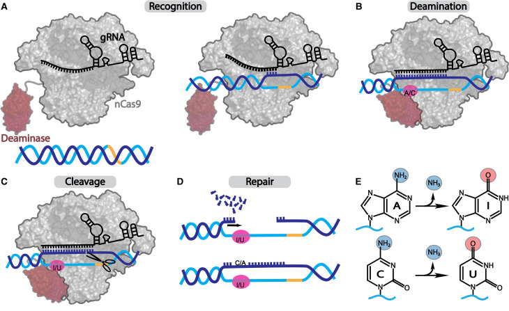
CRISPR-Cas, the bacterial immune systems, have transformed the field of genome editing by providing efficient, easily programmable, and accessible tools for targeted genome editing. DNA base editors (BE) are state-of-the-art CRISPR-based technology, allowing for targeted modifications of individual nucleobases within the genome. Among the BEs, adenine base editors (ABEs) have shown great potential due to their ability to convert A-to-G with high efficiency. However, current ABEs have limitations in terms of their specificity and targeting range. In this review, we provide an overview of the molecular mechanism of ABEs, with a focus on the mechanism of deoxyadenosine deamination by evolved tRNA-specific adenosine deaminase (TadA). We discuss how mutations and adjustments introduced via both directed evolution as well as rational design have improved ABE efficiency and specificity. This review offers insights into the molecular mechanism of ABEs, providing a roadmap for future developments in the precision genome editing field.

Keywords: DNA adenine base editor; DNA base editing; DNA deaminase; deamination; genome editing; tRNA deaminase.


Conflict of interest statement

The authors declare that there are no competing interests associated with the manuscript.

Figures

Figure 1.
Figure 1.. General process of DNA base editing.
DNA base editing is achieved via four events: (A) Recognition: BE RNPs locate appropriate PAM site (yellow) in the genome, unwind the dsDNA to verify DNA complementarity to the gRNA, and if successful, form a stable R-loop; (B) Deamination: the tethered deaminase interacts with the exposed ssDNA bases in the editing window (pink) of the R-loop and catalyzes the deamination reaction; (C) Cleavage: Cas effectors (typically nCas9) cleaves the unedited TS; (D) Repair: the nicked strand in genomic DNA triggers the SSB repair pathway, which uses the edited strand as a template for repair, resulting in the incorporation of the modification into both strands of the DNA. (E) Schematic representation of the deamination reaction catalyzed by ABEs (top, dA-to-dI editing) and CBEs (bottom, dC-to-dU editing).
Figure 2.
Figure 2.. Sequence and structural comparisons of natural tRNA and evolved DNA deaminases.
(A) Alignment of SaTadA, EcTadA, and TadA8e sequences with assigned secondary structure elements on the top (α-helices as gray ovals; β-strands as black arrows). The amino acids mutated during directed evolution are highlighted in red and marked with a circle. Gray boxes signify the substrate-binding loops (L1–L6). The orange box highlights the residues of α5-helix that interact with and stabilize tRNA in the binding pocket of SaTadA and EcTadA. Catalytic residues are highlighted in pink boxes. (B) The substitutions introduced during directed evolution (red spheres) are mapped onto the structure of a single TadA8e domain in complex with the DNA substrate (gray, PDB ID: 6VPC). (C) The aligned structures of EcTadA (orange and yellow, PDB ID: 1Z3A) and TadA8e (red and pink, PDB ID: 6VPC) exhibit an overall similar 3D shape, with the only observed difference being in the conformation of the α5-helix. Both enzymes form homodimers via the same dimerization interface positioning their active sites on opposite sides. The aligned structures of (D) apo EcTadA (orange) and DNA-bound TadA8e (red) and (E) tRNA-bound SaTadA (pale-yellow, PDB ID: 2B3J) and TadA8e (red) showing the position of TadA8e's α5′-helix in respect to the DNA and the tRNA substrates, respectively.
Figure 3.
Figure 3.. Comparison of interactions between TadA variants and their preferred substrates.
(A) In SaTadA (left, pale-yellow and gray, PDB ID: 2B3J), the D104 residue forms a hydrogen bond with the 2′-OH group of U33 ribose. In TadA8e (right, magenta and blue, PDB ID: 6VPC), the N108 residue (equivalent of D104) is positioned closer to the phosphate group and the base of dC−1. (B) Comparison of U33 and dC−1 binding pockets in SaTadA (left) and TadA8e (right), respectively. (C) Comparison of C35 and dC+1 binding pockets in SaTadA (left) and TadA8e (right), respectively. (D) Comparison of SaTadA (left) and TadA8e (right) active sites with the Neb and 8-azaNeb instead of the target adenosine, respectively.

Similar articles

Cited by

References

    1. Landrum, M.J., Lee, J.M., Riley, G.R., Jang, W., Rubinstein, W.S., Church, D.M.et al. (2014) Clinvar: public archive of relationships among sequence variation and human phenotype. Nucleic Acids Res. 42, D980–D985 10.1093/nar/gkt1113 - DOI - PMC - PubMed
    1. Doebley, J.F., Gaut, B.S. and Smith, B.D. (2006) The molecular genetics of crop domestication. Cell 127, 1309–1321 10.1016/j.cell.2006.12.006 - DOI - PubMed
    1. Stafuzza, N.B., Zerlotini, A., Lobo, F.P., Yamagishi, M.E.B., Chud, T.C.S., Caetano, A.R.et al. (2017) Single nucleotide variants and InDels identified from whole-genome re-sequencing of Guzerat, Gyr, Girolando and Holstein cattle breeds. PLoS ONE 12, e0173954 10.1371/journal.pone.0173954 - DOI - PMC - PubMed
    1. Mojica, F.J.M., Díez-Villaseñor, C., García-Martínez, J. and Soria, E. (2005) Intervening sequences of regularly spaced prokaryotic repeats derive from foreign genetic elements. J. Mol. Evol. 60, 174–182 10.1007/s00239-004-0046-3 - DOI - PubMed
    1. Barrangou, R., Fremaux, C., Deveau, H., Richards, M., Boyaval, P., Moineau, S.et al. (2007) CRISPR provides acquired resistance against viruses in prokaryotes. Science 315, 1709–1712 10.1126/science.1138140 - DOI - PubMed

Publication types