. 2023 Aug 25;86(8):1901-1909.
doi: 10.1021/acs.jnatprod.3c00059. Epub 2023 Aug 1.

Flindissone, a Limonoid Isolated from Trichilia prieuriana, Is an LXR Agonist

Affiliations

Flindissone, a Limonoid Isolated from Trichilia prieuriana, Is an LXR Agonist

Mirta Resetar et al. J Nat Prod. .

Abstract

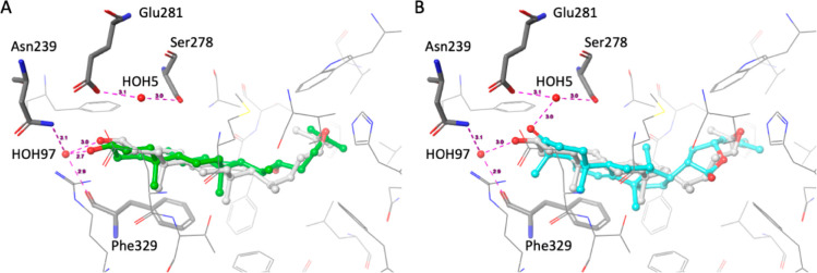
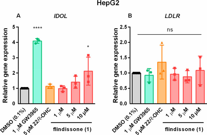
In this study, the ability of six limonoids from Trichilia prieuriana (Meliaceae) to activate the liver X receptor (LXR) was assessed. One of these limonoids, flindissone, was shown to activate LXR by reporter-gene assays. Flindissone is a ring-intact limonoid, structurally similar to sterol-like LXR ligands. In endogenous cellular settings, flindissone showed an activity profile that is characteristic of LXR agonists. It induced cholesterol efflux in THP-1 macrophages by increasing the cholesterol transporter ABCA1 and ABCG1 gene expression. In HepG2 cells, flindissone induced the expression of IDOL, an LXR-target gene that is associated with the downregulation of the LDL receptor. However, unlike synthetic and similarly to sterol-based LXR agonists, flindissone did not induce the expression of the SREBP1c gene, a major transcription factor regulating de novo lipogenesis. Additionally, flindissone also appeared to be able to inhibit post-translational activation of SREBP1c. The results presented here reveal a natural product as a new LXR agonist and point to an additional property of T. prieuriana and other plant extracts containing flindissone.


Conflict of interest statement

The authors declare no competing financial interest.

Figures

Chart 1
Chart 1. Structures of the Compounds Explored in This Study
Figure 1
Figure 1
Concentration–response curves for flindissone (1) and 22R-hydroxycholesterol (22R-OHC) activating the LXRα (A) and LXRβ (B) receptors in luciferase assays employing full-length receptors. Data points show mean ± SEM, N = 4. EC50 and Emax values were obtained by the nonlinear fitting of the log-transformed concentrations using the variable slope. Activation of LXRα-Gal4 (C) and LXRβ-Gal4 (D). Bars show mean, error bars SD, and data points individual values. N = 3. One-way ANOVA with Dunnett’s post hoc test. Significance indicated in comparison to the DMSO (0.1%) control: *: p = 0.05, **: p = 0.01, ***: p = 0.001, ****: p = 0.0001, no indication = not significant.
Figure 2
Figure 2
Depiction of the likely binding mode of (A) 22R-hydroxycholesterol (22R-OHC; green carbon atoms) and (B) flindissone (1; cyan carbon atoms) for the LXRβ ligand binding domain, derived by docking. 24(S),25-Epoxycholesterol (eCH), the ligand present in the cocrystal structure used for docking (PDB 1P8B), is depicted with gray carbon atoms. Magenta dashed lines indicate hydrogen bond interactions; the numbers indicate distances in Å. Amino acid residues forming hydrogen bonds with the ligands via H2O molecules are marked by the thick tube representation.
Figure 3
Figure 3
Flindissone (1)-mediated stimulation of cholesterol efflux in THP-1 M0 macrophages (A). The gene expression (mRNA level) of the cholesterol transporter ABCA1, measured by RT-qPCR (B) and its protein quantification with the representative blot below (C). Upper panel: 250 kDa: ABCA1. Lower panel: 50 kDa: α/β-tubulin. For all graphs: bars show mean, error bars SD, and data points individual values. N = 3. One-way ANOVA with Dunnett’s post hoc test. Significance indicated in comparison to the DMSO (0.1%) control: *: p = 0.05, **: p = 0.01, ***: p = 0.001, ****: p = 0.0001, no indication = not significant.
Figure 4
Figure 4
Flindissone-mediated target-gene regulation in the HepG2 cell line. The mRNA levels of IDOL (A) and LDLR (B) were measured by RT-qPCR. N = 3. Bars show mean ± SD. One-way ANOVA with Dunnett’s post hoc test. Significance indicated in comparison to the DMSO (0.1%) control: *: p = 0.05, ****: p = 0.0001, ns = not significant.
Figure 5
Figure 5
Flindissone-mediated regulation of lipogenesis in HepG2 cells. Measurements of the cellular neutral lipid content by Oil Red O staining (A). N = 4. Representative images were taken at 40× magnification. Gene expression (mRNA) of SREBF1 (B), measured by RT-qPCR. N = 3. Quantification of the SREBP1c 130 kDa (C) and cleaved 60 kDa product relative to its 130 kDa precursor (D). Representative blots of the SREBP1c protein levels below. Upper panel: 130 kDa: SREBP1c precursor; 60 kDa: cleaved SREBP1c. Lower panel: 42 kDa: actin. For all graphs: Bars show mean, error bars SD, and data points individual values. One-way ANOVA with Dunnett’s post hoc test. Significance indicated in comparison to the DMSO (0.1%) control: *: p = 0.05, **, p = 0.01, no indication or ns = not significant.

References

    1. Lemmens R. H. M.Trichilia Prieureana A. Juss. In PROTA (Plant Resources of Tropical Africa/Ressources végétales de l’Afrique tropicale); Louppe D.; Oteng-Amoako A. A.; Brink M., Eds.; Wageningen, Netherlands, 2008.
    1. Luo J.; Sun Y.; Li Q.; Kong L. Nat. Prod. Rep. 2022, 39, 1325–1365. 10.1039/D2NP00015F. - DOI - PubMed
    1. Passos M. S.; Nogueira T. S. R.; Azevedo O. de A.; Vieira M. G. C.; Terra W. da S.; Braz-Filho R.; Vieira I. J. C Phytochem. Rev. 2021, 20, 1055–1086. 10.1007/s11101-020-09737-x. - DOI
    1. Pagna J. I. M.; Mbekou I. M. K.; Tsamo A. T.; Mkounga P.; Frese M.; Stammler H.-G.; Fekam F. B.; Lenta B. N.; Sewald N.; Nkengfack A. E. Zeitschrift für Naturforsch. B 2021, 76, 439–446. 10.1515/znb-2021-0057. - DOI
    1. Wang B.; Tontonoz P. Nat. Rev. Endocrinol. 2018, 14, 452–463. 10.1038/s41574-018-0037-x. - DOI - PMC - PubMed

Publication types

MeSH terms

LinkOut - more resources