. 2023 Sep;621(7979):627-634.
doi: 10.1038/s41586-023-06477-8. Epub 2023 Aug 1.

Central role of Tim17 in mitochondrial presequence protein translocation

Affiliations

Central role of Tim17 in mitochondrial presequence protein translocation

Laura F Fielden et al. Nature. 2023 Sep.

Abstract

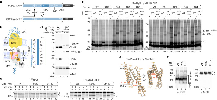
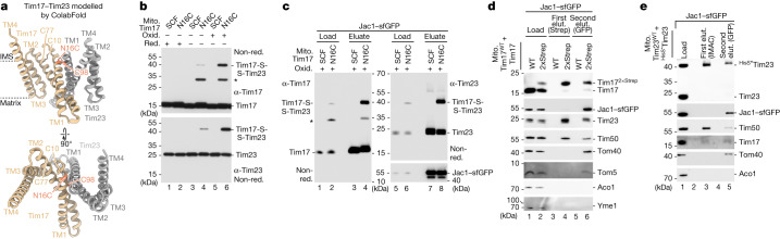
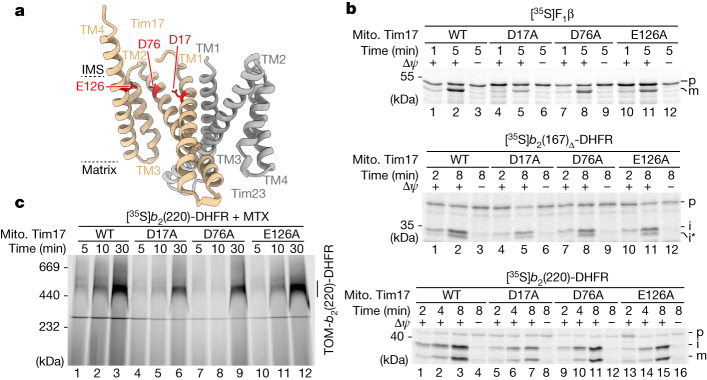
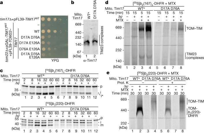
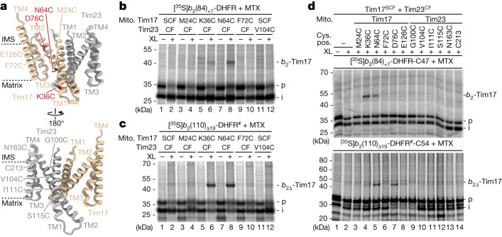
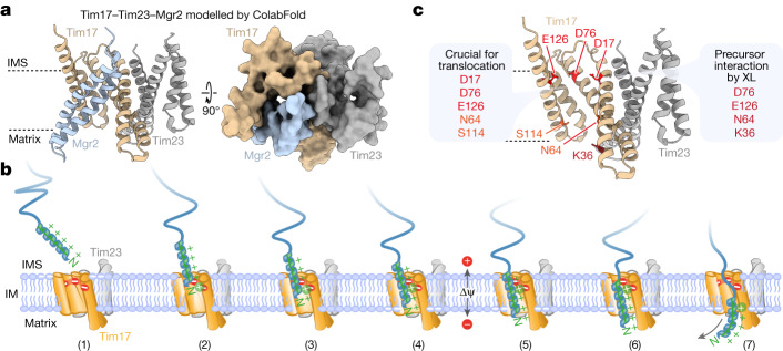
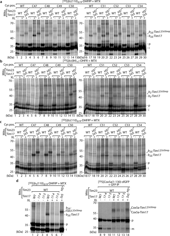
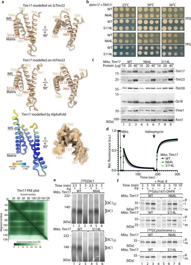
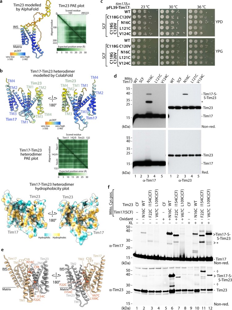
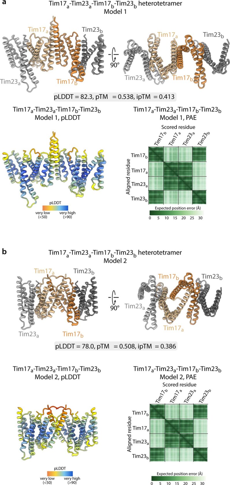
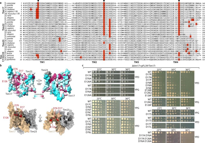
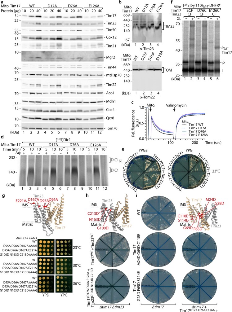
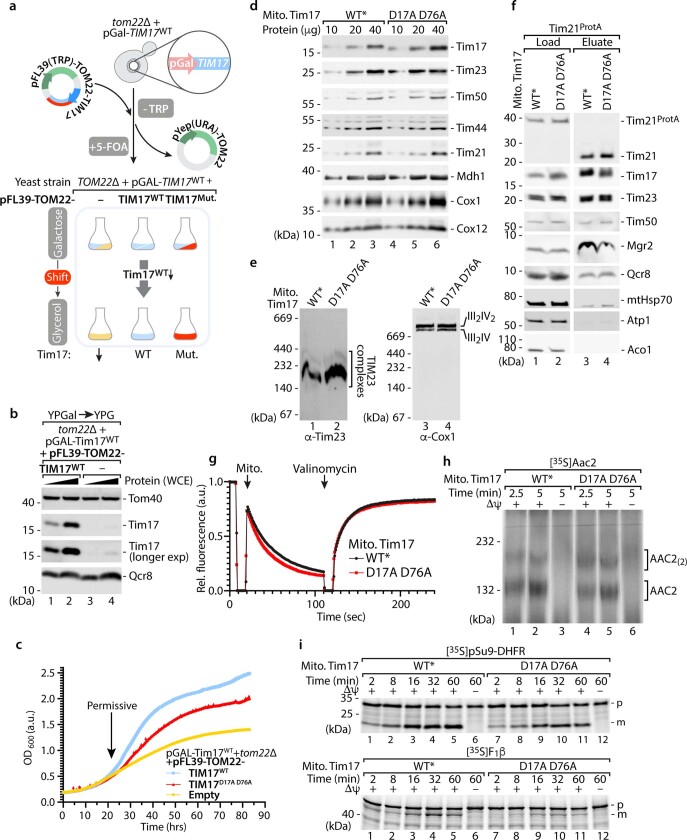
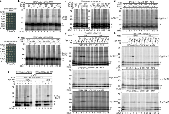
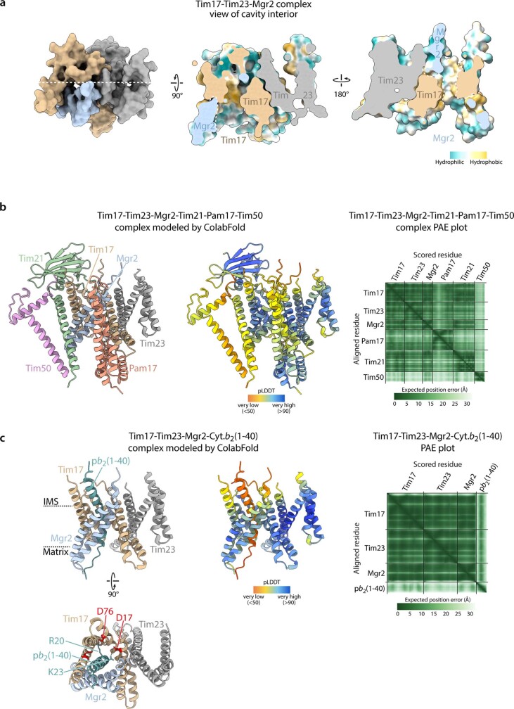
The presequence translocase of the mitochondrial inner membrane (TIM23) represents the major route for the import of nuclear-encoded proteins into mitochondria1,2. About 60% of more than 1,000 different mitochondrial proteins are synthesized with amino-terminal targeting signals, termed presequences, which form positively charged amphiphilic α-helices3,4. TIM23 sorts the presequence proteins into the inner membrane or matrix. Various views, including regulatory and coupling functions, have been reported on the essential TIM23 subunit Tim17 (refs. 5-7). Here we mapped the interaction of Tim17 with matrix-targeted and inner membrane-sorted preproteins during translocation in the native membrane environment. We show that Tim17 contains conserved negative charges close to the intermembrane space side of the bilayer, which are essential to initiate presequence protein translocation along a distinct transmembrane cavity of Tim17 for both classes of preproteins. The amphiphilic character of mitochondrial presequences directly matches this Tim17-dependent translocation mechanism. This mechanism permits direct lateral release of transmembrane segments of inner membrane-sorted precursors into the inner membrane.


Conflict of interest statement

The authors declare no competing interests.

Figures

Fig. 1
Fig. 1. Matrix-targeted and inner membrane-sorted precursors interact with Tim17.
a, Schematic of preprotein constructs of identical length. Both preproteins are derivatives of cytochrome b2 (containing an N-terminal matrix-targeting signal (Matrix targ.)) fused to DHFR. The b2(84)+7-DHFR contains an inner membrane sorting signal (Sort.) containing a transmembrane domain and a seven amino acid linker (+7). The b2(110)Δ19-DHFR is imported into the mitochondrial matrix because the sorting signal (19 amino acids) is deleted (Δ). IMP, inner membrane protease. b, Model of arrested b2-DHFR precursor proteins depicted in a. Cyt., cytosol; IM, inner membrane; IMS, intermembrane space; OM, outer membrane; Pos., position; TIM23, presequence translocase of the inner membrane; PAM, presequence translocase-associated motor; Δψ, membrane potential. c, Import of 35S-labelled b2(84)+7-DHFR with cysteine residues at the indicated positions (pos.) in wild-type (WT) or Tim172xStrep (2xS) and HisSUMOstarTim23 (HS*) mitochondria in the presence of MTX followed by chemical crosslinking with MBS (XL). Samples were analysed by SDS–PAGE and autoradiography. b2-Tim17, b2(84)+7-DHFR-Tim17 crosslinking product; i, intermediate; p, precursor. d, Immunodecoration depicting the mass shift of tagged Tim172xStrep and HisSUMOstarTim23. e, AlphaFold model of full-length Tim17 (Saccharomyces cerevisiae; AF-P39515). Model shown in ribbon representation is viewed parallel to the membrane (left) and from the IMS (right). Dashed lines indicate the predicted membrane region. Orange, asparagine 64 (N64), serine 114 (S114) residues. f, Protein complexes of isolated wild-type Tim17 and mutant yeast mitochondria were analysed by Blue Native (BN)–PAGE and immunodecoration. g, Import of radiolabelled matrix precursor proteins pSu9-DHFR (pSu9, presequence of Neurospora crassa ATP synthase subunit 9) and ATP synthase subunit 2 (Atp2), also named F1β, into isolated wild-type Tim17 and mutant yeast mitochondria followed by SDS–PAGE and autoradiography. m, mature.
Fig. 2
Fig. 2. Lateral transmembrane cavities face toward opposite sides of the Tim17–Tim23 dimer.
a, ColabFold structural protein complex model of Tim17 (tan) and Tim23 (grey) heterodimers. Tan, endogenous intramolecular disulfide bond formed between Tim17 Cys10 (C10) and Cys77 (C77); orange, Tim17 Cys16 (N16C) and Tim23 Cys98 (C98) forming an intermolecular disulfide bond. b, In organello disulfide bond formation between Tim17 and Tim23. Mitochondria isolated from yeast strains expressing SCF Tim17 or Tim17 N16C were left untreated or treated with the reductant dithiothreitol (DTT; red) or oxidant 4,4′-dipyridyl disulfide (4-DPS; Oxid.) followed by SDS–PAGE. Tim17-S-S-Tim23, disulfide-linked Tim17 and Tim23. *Unidentified Tim17-specific conjugate. c, Import of Jac1–sfGFP into SCF Tim17 or N16C mitochondria followed by oxidation with 4-DPS. Jac1–sfGFP-containing TOM–TIM23 supercomplexes were isolated with a GFP nanobody. Load (1%) and eluate (100%) fractions were analysed by SDS–PAGE and immunodecoration. d,e, Import of Jac1–sfGFP into wild-type or Tim172xStrep (d) and HisSumo*Tim23 mitochondria (e). TIM23 complexes were isolated by tandem purification first using streptavidin (Strep; d) or immobilized metal affinity chromatography (IMAC; e) followed by purification of Jac1–sfGFP-containing TOM–TIM23 supercomplexes using a GFP nanobody. Fractions were analysed by SDS–PAGE and immunodecoration. Load, 0.25%; first elut., first elution (2.5%); second elut., second elution (100%).
Fig. 3
Fig. 3. Negative charges of Tim17 transmembrane domains are crucial for presequence protein translocation.
a, ColabFold model of S. cerevisiae Tim17 (tan) and Tim23 (grey) heterodimer showing, as red sticks, the locations of negatively charged residues aspartic acid 17 (D17) and 76 (D76) and glutamic acid 126 (E126) within the transmembrane region of Tim17. b, Import of radiolabelled F1β (matrix), b2(167)Δ-DHFR (matrix) and b2(220)-DHFR (inner membrane-sorted) precursors into isolated mitochondria; subsequently, they were treated with proteinase K and subjected to SDS–PAGE and autoradiography. i*, intermediate. c, MTX preincubated radiolabelled b2(220)-DHFR was imported into isolated wild-type Tim17 or mutant mitochondria followed by BN–PAGE and autoradiography. TOM-b2(220)-DHFR, MPP and IMP processed b2(220)-DHFR was stabilized at the TOM complex.
Fig. 4
Fig. 4. Tim17-dependent presequence translocation is initiated at the negative charged patch.
a, Growth analysis of yeast strains expressing wild-type or tim17 variants with the indicated double or triple negative charge mutation on non-fermentable glycerol (YPG) medium at 19 °C. b, Mitochondrial protein complexes were analysed by BN–PAGE as described in Fig. 1f. WT*, mitochondria isolated from tom22Δ, pGal-TIM17WT + pFL39-TOM22 yeast cells with levels of Tim17 comparable with Tim17D17A_D76A mutant mitochondria. c, Protein import into wild-type and Tim17D17A_D76A mitochondria was analysed as described for Fig. 3c.d, Radiolabelled b2(167)Δ-DHFR was imported and accumulated in import sites of isolated mitochondria in the presence of MTX as indicated. Samples were analysed by BN–PAGE and autoradiography. TOM–TIM, TOM–TIM23 preprotein stabilized supercomplex. e, Accumulation of radiolabelled b2(220)-DHFR in TOM–TIM23 import sites of isolated mitochondria as in Fig. 3c, followed by treatment with proteinase (Prot.) K.
Fig. 5
Fig. 5. Arrested precursor proteins are associated throughout the lateral cavity of Tim17.
a, ColabFold protein complex model of S. cerevisiae Tim17 (tan) and Tim23 (grey) heterodimer showing the location of cysteine mutants (tan/dark red/grey) within the transmembrane regions of Tim17 (top) or Tim23 (bottom). b,c, Radiolabelled MTX-stabilized b2(84)+7-DHFR (b) and b2(110)Δ19-DHFR (c) constructs were accumulated in mitochondria with cysteine residues in Tim17 (SCF) or Tim23 (CF) at TOM–TIM23 import sites. Subsequently, samples were chemically crosslinked with MBS (XL) and analysed by SDS–PAGE and autoradiography. b2∆-Tim17, b2(110)∆19-DHFR-Tim17 crosslinking product. d, b2-DHFR constructs containing a cysteine at position 47 (C47; b2(84)+7-DHFR) or 54 (C54; b2(110)∆19-DHFR) were analysed as described in c but chemically crosslinked with BMOE (XL).
Fig. 6
Fig. 6. Model for mitochondrial presequence translocation across the inner mitochondrial membrane.
a, Structural protein complex model of the interaction between Tim17 (tan), Mgr2 (blue) and Tim23 (grey) generated by ColabFold, shown as cartoon (left) and surface (right) representations. b, The Tim17 TIS composed of the acidic patch of the lateral transmembrane cavity of Tim17 is positioned close to the intermembrane space side and attracts the positive charge of the N terminus of the precursor protein (1). The hydrophobic residues of the amphiphilic presequence engage with the outer leaflet of the lipid bilayer, which stabilizes the α-helical conformation of the presequence and reinforces the interaction between the presequence and the TIS (2). Successive positively charged residues on the hydrophilic side of the α-helical presequence enable insertion of the hydrophobic face into the lipid bilayer in steps of single turns (3 and 4). The negatively charged phospholipid head groups on the matrix face of the inner membrane attract the positively charged amino terminus (5). The membrane potential (Δψ) and following positive residues support the emergence of the presequence on the matrix side (6) and engagement with the mitochondrial presequence translocase-associated motor, which aids the remaining steps of import into the matrix (7). c, Summary of the findings of the current study depicted onto the ColabFold structural model of the interaction between Tim17 (tan) and Tim23 (grey). Red, residues of the Tim17 TIS; orange, hydrophilic residues; dark red, residues crosslinked to arrested precursor protein intermediates.
Extended Data Fig. 1
Extended Data Fig. 1. Arrested presequence precursors are in proximity to Tim17.
ac, Import of 35S-labelled b2(110)∆19-DHFR# and b2(84)+7-DHFR with cysteine residues at the indicated positions (Cys pos.) in wildtype (WT) or Tim172xStrep (2xS) and HisSUMOstarTim23 (HS*) mitochondria in the presence of methotrexate (MTX) followed by chemical crosslinking (XL) with m-maleimidobenzoyl-N-hydroxysuccinimide ester (MBS, a) or bismaleimidoethane (BMOE, b and c). Samples were analysed by SDS-PAGE and autoradiography. b2∆-Tim17, b2(110)∆19-DHFR#-Tim17 crosslinking product; b2∆-Tim172xStrep, b2(110)∆19-DHFR#-Tim172xStrep crosslinking product; b2-Tim17, b2(84)+7-DHFR-Tim17 crosslinking product; b2-Tim172xStrep, b2(84)+7-DHFR-Tim172xStrep crosslinking product; p, precursor; i, intermediate. d, Radiolabelled b2(110)∆19-DHFR# (lanes 1–7) or Cox5a(1–130)-sfGFP (lanes 8–14) were imported into Tim17 WT, tim17-4, tim17-5 or Tim172xStrep (2xS) and HisSumo*Tim23 (HS*) mitochondria following heat shock. Crosslinked products were either directly analysed (b2(110)∆19-DHFR#, lanes 1–7) or purified using a GFP nanobody (Cox5a-sfGFP, lanes 8–14) prior to analysis by SDS-PAGE and autoradiography. Cox5a-Tim17, Cox5a-sfGFP-Tim17 crosslinking product; Cox5a-Tim172xStrep, Cox5a-sfGFP-Tim172xStrep crosslinking product; m, mature.
Extended Data Fig. 2
Extended Data Fig. 2. Hydrophilic residues within the lateral Tim17 cavity are crucial for matrix translocation.
a, Tim17 (S. cerevisiae) modelled on the Tim22 (S.c.) structure (top; PDB ID 6LO8) and Tim22 (H. sapiens) structure (middle; PDB ID 7CGP). Dashed lines indicate the transmembrane (TM) segments of Tim17 (orange, asparagine 64 (N64), and serine (S114) residues). Predicted Local Distance Difference Test (pLDDT) score together with predicted aligned error plot (PAE) as well as surface representation of Tim17 as seen from the intermembrane space of the Tim17 AlphaFold model depicted in Fig. 1e (bottom). IMS, intermembrane space. b, Growth analysis of yeast strains expressing WT, Tim17N64L or Tim17S114L variants on agar with fermentable (dextrose/glucose, YPD) and non-fermentable (glycerol, YPG) medium at the indicated temperatures. c, Protein amounts of WT, Tim17N64L and Tim17S114L mitochondria isolated from yeast cells grown on non-fermentable media (YPG) at 23 °C, analysed by SDS-PAGE and immunodecoration against the indicated antibodies. d, Membrane potential assessment of isolated WT, Tim17N64L and Tim17S114L mitochondria (from yeast cells grown in YPG at 23 °C). The membrane potential was assessed by fluorescence quenching using the potential sensitive dye 3,3’-dipropylthiadicarbocyanine iodide and subsequently dissipated by addition of valinomycin. e, Import of radiolabelled metabolite carrier protein dicarboxylate carrier 1 (Dic1) into isolated WT, Tim17N64L and Tim17S114L mitochondria, followed by blue native PAGE and autoradiography. Δψ, membrane potential; DIC1(2), assembled Dic(oligomer). f, Import of radiolabelled b2(167)-DHFR and cytochrome c1 into isolated WT, Tim17N64L and Tim17S114L mitochondria followed by SDS-PAGE and autoradiography. Source data
Extended Data Fig. 3
Extended Data Fig. 3. Analysis of the Tim17-Tim23 back-to-back interaction with lateral cavities on opposing sides of the heterodimer.
a, Predicted Local Distance Difference Test (pLDDT) score plotted onto the Tim23 AlphaFold model (AF-P32897) alongside the associated predicted aligned error plot (PAE). b, Tim17-Tim23 heterodimer modelled by ColabFold. pLDDT score (upper), predicted aligned error (PAE, middle) and surface structure hydrophobicity (lower) plots. c, Growth analysis of yeast strains expressing WT or Tim17 cysteine mutant variants in the semi-cysteine-free background (SCF) as described for Extended Data Fig. 2b. d, Analysis of mitochondria isolated from yeast strains expressing WT, semi-cysteine-free Tim17 (SCF) and Tim17 cysteine variants by non-reducing (Non-red.) and reducing (Red.) SDS-PAGE and immunodecoration with the indicated antibodies. Tim17, Tim17 monomer; Tim23, Tim23 monomer; Tim17-S-S-Tim23, disulfide-linked Tim17 and Tim23; *, unidentified Tim17-specific conjugate e, ColabFold structural protein complex model of Tim17 (tan) and Tim23 (grey) heterodimer showing location of cysteine mutants (orange) at the dimer interface of the transmembrane regions of Tim17 and Tim23. Dotted lines, formation of disulphide bond or crosslinking product. f, Disulfide bond or crosslink formation between Tim17 and Tim23. Mitochondria isolated from yeast strains expressing Tim17 or Tim23 cysteine mutant variants in a semi-cysteine-free Tim17 (SCF) and Tim23 WT or cysteine-free (CF) background were left untreated or treated with oxidant (4-DPS; Oxidant) or crosslinker (BMOE; XL) followed by non-reducing SDS-PAGE and immunodecoration. ♦, unidentified Tim23-specific conjugate.
Extended Data Fig. 4
Extended Data Fig. 4. Structural modelling of Tim17-Tim23 heterotetramers.
a,b, Representative ColabFold structural protein complex models of Tim17 (a/b: tan/orange) and Tim23 (a/b: grey/dark grey) within putative weakly predicted heterotetrameric arrangements with associated pLDDT scores, PAE plots, predicted TM score (pTM) and interface pTM (ipTM) scores. These two distinct models predicted by ColabFold represent those with the highest ranking.
Extended Data Fig. 5
Extended Data Fig. 5. Sequence alignment of Tim17 protein family with conserved negative charges within the TMDs and growth phenotype of corresponding tim17 mutants.
a, Sequence alignment of Tim17 from Saccharomyces cerevisiae, Schizosaccharomyces pombe, Neurospora crassa, Caenorhabditis elegans, Drosophila melongaster, Danio rerio, Homo sapiens, Mus musculus and Arabidopsis thaliana (paralog 1) generated by TMcoffee. Predicted TMDs were assigned following sequence alignment of S. c. Tim17 with S. c. Tim22 and according to the cyro-EM structure of Tim22 (6LO8). Grey boxes, predicted transmembrane (TM) domains; Red, highly conserved negatively charged residues within predicted TM domains; Arrowheads, negatively charged residues analysed in this study. b, Tim17 and Tim23 amino acid sequence conservation mapped onto the surface of the ColabFold predicted Tim17-Tim23 heterodimer (upper panel). Space filling model of the Tim17-Tim23 heterodimer with the conserved negatively charged acidic patch formed by D17, D76 and E126 indicated (lower panel). c, Growth analysis of WT and tim17 mutants in tim17Δ background related to Extended Data Table 1. Yeast strains expressing WT or the indicated tim17 mutant variant were grown on non-fermentable (glycerol, YPG) or fermentable (dextrose/glucose, YPD) agar medium at the indicated temperatures.
Extended Data Fig. 6
Extended Data Fig. 6. Tim17D17A and Tim17D76A mitochondria have no obvious secondary defects and introduction of equivalent negative charges into Tim23 or lower parts of the lateral cavity of Tim17.
a, Protein amounts of WT, Tim17D17A, Tim17D76A and Tim17E126A mitochondria isolated from yeast cells grown in YPG at 23 °C, followed by SDS-PAGE and immunodecoration with the indicated antibodies. b, Protein complexes of isolated Tim17 WT and mutant yeast mitochondria analysed by BN-PAGE and immunodecoration with the indicated antibodies. TIM23, presequence translocase of the inner membrane; TOM, translocase of the outer mitochondrial membrane. c, Assessment of the mitochondrial membrane potential of isolated mitochondria as described in Extended Data Fig. 2d. d, Import of radiolabelled metabolite carrier protein Dic1 into isolated WT and tim17 mutant mitochondria, followed by BN-PAGE and autoradiography. e, Growth analysis of tim17 tim23 mutant yeast strains expressing Tim17SCF+Tim23CF or Tim17D17C+Tim23CF on agar media containing galactose (YPGal) and glycerol (YPG) at 23 °C. f, Import of 35S-labelled b2(110)∆19-DHFR# into Tim17 WT or cysteine mutant mitochondria in the presence of MTX followed by chemical crosslinking (MBS; XL), SDS-PAGE and autoradiography. g, ColabFold structural protein complex model of S. c. Tim17 (tan) and Tim23 (grey) heterodimer with the location of the negatively charged residues (red) within the transmembrane domains and adjacent segments of Tim23 (upper panel). Growth analysis of tim23 mutant yeast strains expressing Tim23 WT or negative charge mutant variants as for Extended Data Fig. 2b (lower panel). AAA, Tim23 D95A D96A D167A. h, i, ColabFold structural protein complex model as for g with the location of residues equivalent to Tim17 D17, D76 and E126 that were introduced into the transmembrane regions of Tim23 (h) or locations with equivalent charged residues, located lower within the lateral cavity of Tim17, were introduced (i) (red; upper panels). Growth phenotypes of tim17 tim23 mutant yeast strains in the tim17∆tim23∆ background (h) or tim17 mutants in the tim17∆ and tim17∆, Tim17D17A_D76A_E126A background (i) on 5-FOA medium at 23 °C (lower panel). Source data
Extended Data Fig. 7
Extended Data Fig. 7. Yeast expressing Tim17D17A_D76A and analysis of Tim17D17A_D76A mitochondria.
a, Graphical depiction of the approach to generate Tim17 mutants by placing chromosomal TIM17 under the control of the galactose promotor (pGAL-TIM17) in a TOM22 deletion background. This strain was transformed with single copy pFL(TRP) plasmids, encoding WT TOM22 and the desired TIM17 alleles. After elimination of the pYEP(URA)-TOM22 cover plasmid by 5-Fluoroorotic acid (5-FOA) treatment, the pFL plasmid is maintained, as the yeast cells require the TOM22 gene for viability. pGal, galactose inducible promoter; TRP, tryptophan; URA, uracil; Mut., tim17 mutant. b, Analysis of Tim17WT protein depletion by shifting the pGAL-TIM17WT strain from galactose to fermentable glycerol medium for 21 h. Protein levels in yeast whole cell extracts (WCE) were analysed by SDS-PAGE and immunodecoration. c, Growth analysis of tom22Δ, pGal-TIM17WT + pFL39-TOM22 yeast strains expressing Tim17WT (blue), Tim17D17A_D76A (red) or no Tim17/‘empty’ (yellow) in glycerol-containing media at 25 °C. Arrow indicates the time before the growth of the strain expressing Tim17D17A_D76A is affected compared to the WT strain. d, Protein amounts of WT* and Tim17D17A_D76A mitochondria isolated from yeast cells shifted to non-fermentable media (YPG) at 23 °C, analysed by SDS-PAGE and immunodecoration against the indicated antibodies. WT*, WT strain with Tim17 protein levels comparable to Tim17D17A_D76A mutant e, Protein complexes of isolated Tim17 WT and Tim17D17A_D76A mutant yeast mitochondria were analysed by BN-PAGE and immunodecoration. III2IV, III2IV2, respiratory chain supercomplexes. f, TIM23 complex isolation from digitonin-solubilised Tim17WT + Tim21ProtA or Tim17D17A_D76A + Tim21ProtA mitochondria. Bound protein complexes were analysed by SDS-PAGE and immunodecoration with the indicated antibodies. ProtA, Protein A; Load, 1%; Eluate 100%. g, Mitochondrial membrane potential of WT and Tim17D17A_D76A mitochondria isolated from yeast cells grown in non-fermentable glycerol medium. Assessment of the membrane potential of isolated mitochondria as described in Extended Data Fig. 2d. h, Import of radiolabelled metabolite carrier ADP/ATP carrier (Aac2) into isolated WT and Tim17D17A_D76A mitochondria followed by BN-PAGE and autoradiography. AAC(2), assembled ADP/ATP carrier(oligomer). i, Import of radiolabelled pSu9-DHFR (top) and F1β (Atp2, bottom) into WT and Tim17D17A_D76A mitochondria isolated as described in Extended Data Fig. 2f. Source data
Extended Data Fig. 8
Extended Data Fig. 8. Interaction of precursor proteins with the lateral Tim17 cavity is membrane potential-dependent.
a, Growth phenotypes of the indicated tim17 (top panel) or tim23 (bottom panel) cysteine mutants in the tim17∆tim23∆ background on YPG agar at 23 °C. b-e, Arrest of 35S-labelled Cox5a(1–130)-sfGFP (b and e), b2(84)+7-DHFR (c) or b2(110)∆19-DHFR# (d) into Tim17SCF and Tim23CF mitochondria with cysteine residues introduced at the indicated positions. Chemical crosslinking (XL) was performed with MBS followed by SDS-PAGE and autoradiography. f, Methotrexate (MTX) preincubated, radiolabelled b2(84)+7-DHFR or b2(110)∆19-DHFR# was imported into Tim17SCF or Tim17SCF_N16C mitochondria in the presence or absence of a membrane potential (∆ψ) across the inner membrane followed by chemical crosslinking (XL) with MBS. Samples were subsequently analysed by SDS-PAGE and autoradiography. g,h, Radiolabelled b2(84)+7-DHFR (g) or b2(110)∆19-DHFR# (h) constructs with cysteine residues at the indicated positions were imported into Tim17SCF+Tim23CF control or mutant mitochondria with cysteine residues at the indicated position (Cys. pos.) within the transmembrane cavity. Import was followed by chemical crosslinking (XL) with BMOE, SDS-PAGE and autoradiography.
Extended Data Fig. 9
Extended Data Fig. 9. TIM23 complex modelling.
a, ColabFold structural protein complex prediction of the interaction between S. c. Tim17, Tim23 and Mgr2 as depicted in Fig. 6a (left) and displayed ‘cut-open’ with hydrophilic and hydrophobic residues mapped onto the protein surface (middle and right). b, ColabFold structural prediction of the complex formed by S. c. Tim17 (tan), Tim23 (grey), Mgr2 (blue), Pam17 (salmon), Tim21 (green) and Tim50 (pink) with associated pLDDT scores and PAE plots. The TIM23 subunits with transmembrane segments Mgr2, Pam17, Tim21 and Tim50 are all predicted to associate at the Tim17 side of the Tim17-Tim23 heterodimer. Compared to the high confidence prediction that Mgr2 can directly associate to the Tim17 lateral cavity; all the other subunits have lower confidence predictions and are more peripheral compared to Mgr2. c, ColabFold structural protein complex prediction of the heterotrimer formed by S. c. Tim17 (tan), Tim23 (grey), Mgr2 (blue) and residues 1–40 of cytochrome b2 (pb2) including the presequence with associated pLDDT scores and PAE plots. Dotted lines, putative interactions between Tim17 D17 and D76 and indicated positively charged residues of pb2; red, Tim17 TIS residues.

References

    1. Neupert W, Herrmann JM. Translocation of proteins into mitochondria. Annu. Rev. Biochem. 2007;76:723–749. doi: 10.1146/annurev.biochem.76.052705.163409. - DOI - PubMed
    1. Wiedemann N, Pfanner N. Mitochondrial machineries for protein import and assembly. Annu. Rev. Biochem. 2017;86:685–714. doi: 10.1146/annurev-biochem-060815-014352. - DOI - PubMed
    1. Vögtle F-N, et al. Global analysis of the mitochondrial N-proteome identifies a processing peptidase critical for protein stability. Cell. 2009;139:428–439. doi: 10.1016/j.cell.2009.07.045. - DOI - PubMed
    1. Morgenstern M, et al. Quantitative high-confidence human mitochondrial proteome and its dynamics in cellular context. Cell Metab. 2021;33:2464–2483.e18. doi: 10.1016/j.cmet.2021.11.001. - DOI - PMC - PubMed
    1. Schulz C, Schendzielorz A, Rehling P. Unlocking the presequence import pathway. Trends Cell Biol. 2015;25:265–275. doi: 10.1016/j.tcb.2014.12.001. - DOI - PubMed

Publication types

MeSH terms

Substances

LinkOut - more resources