. 2023 Aug 1;10(5):e200144.
doi: 10.1212/NXI.0000000000200144. Print 2023 Sep.

Structural Changes of Cutaneous Immune Cells in Patients With Type 1 Diabetes and Their Relationship With Diabetic Polyneuropathy

Affiliations

Structural Changes of Cutaneous Immune Cells in Patients With Type 1 Diabetes and Their Relationship With Diabetic Polyneuropathy

Xiaoli Hu et al. Neurol Neuroimmunol Neuroinflamm. .

Erratum in

  • Missing Full Disclosures.
    [No authors listed] [No authors listed] Neurol Neuroimmunol Neuroinflamm. 2025 Jan;12(1):e200342. doi: 10.1212/NXI.0000000000200342. Epub 2024 Oct 30. Neurol Neuroimmunol Neuroinflamm. 2025. PMID: 39475708 Free PMC article. No abstract available.

Abstract

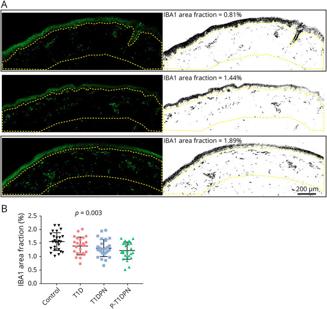
Background and objectives: Diabetic polyneuropathy (DPN) is a complication of diabetes characterized by pain or lack of peripheral sensation, but the underlying mechanisms are not yet fully understood. Recent evidence showed increased cutaneous macrophage infiltration in patients with type 2 diabetes and painful DPN, and this study aimed to understand whether the same applies to type 1 diabetes.

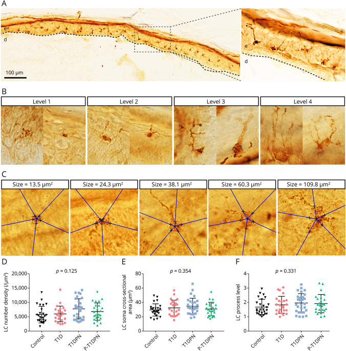
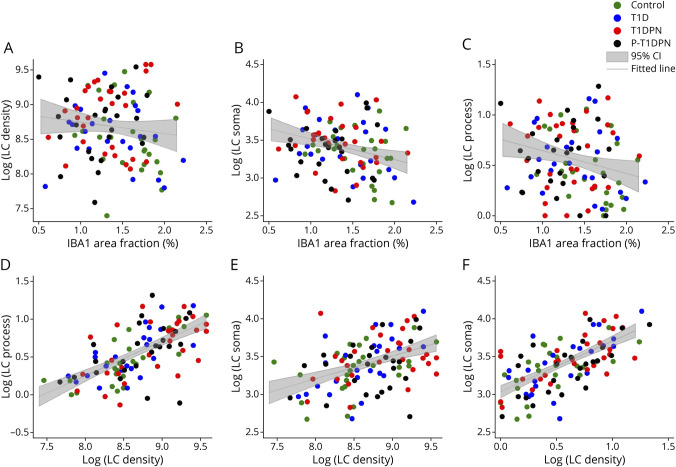
Methods: The study included 104 participants: 26 healthy controls and 78 participants with type 1 diabetes (participants without DPN [n = 24], participants with painless DPN [n = 29], and participants with painful DPN [n = 25]). Two immune cells, dermal IBA1+ macrophages and epidermal Langerhans cells (LCs, CD207+), were visualized and quantified using immunohistological labeling and stereological counting methods on skin biopsies from the participants. The IBA1+ macrophage infiltration, LC number density, LC soma cross-sectional area, and LC processes were measured in this study.

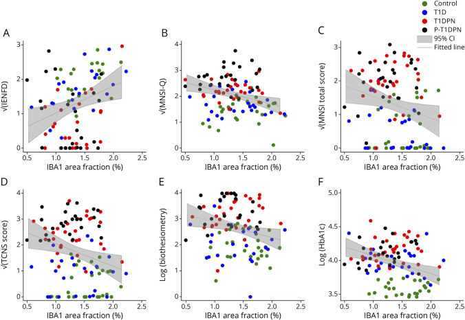
Results: Significant difference in IBA1+ macrophage expression was seen between the groups (p = 0.003), with lower expression of IBA1 in participants with DPN. No differences in LC morphologies (LC number density, soma cross-sectional area, and process level) were found between the groups (all p > 0.05). In addition, IBA1+ macrophages, but not LCs, correlated with intraepidermal nerve fiber density, Michigan neuropathy symptom inventory, (questionnaire and total score), severity of neuropathy as assessed by the Toronto clinical neuropathy score, and vibration detection threshold in the whole study cohort.

Discussion: This study showed expressional differences of cutaneous IBA1+ macrophages but not LC in participants with type 1 diabetes-induced DPN compared with those in controls. The study suggests that a reduction in macrophages may play a role in the development and progression of autoimmune-induced diabetic neuropathy.


Conflict of interest statement

P. Karlsson has received personal fees from Grünenthal, Alnylam, and Vertex Pharmaceuticals and has received a research grant from Merck outside the submitted work. Other authors have no conflicts of interest to state. Go to Neurology.org/NN for full disclosures.

Figures

Figure 1
Figure 1. Immunofluorescent Labeling and Quantification of IBA1+ Macrophages in Human Skin Sections
(A) Representative images of different expression levels of IBA1+ macrophages in skin sections. (B) The quantification of IBA1 area fraction in dermis. Enclosed area by yellow dotted line: the region of interest (dermis that was 300 μm below epidermis-dermis border); T1D = type 1 diabetes without diabetic polyneuropathy; T1DPN = type 1 diabetes with painless diabetic polyneuropathy; P-T1DPN = type 1 diabetes with painful diabetic polyneuropathy.
Figure 2
Figure 2. Quantification of Cutaneous Langerhans Cell Morphologies
(A) Representative image of Langerhans cells in healthy human epidermis. (B) Representative structures of 4 Langerhans cell process levels. (C) Representative images of Langerhans cell soma cross-sectional area estimation. (D) The quantification of Langerhans cell number density in epidermis. (E) The quantification of Langerhans cell soma cross-sectional area in epidermis. (F) The quantification of Langerhans cell process levels in epidermis. LC = Langerhans cell; T1D = type 1 diabetes without diabetic polyneuropathy; T1DPN = type 1 diabetes with painless diabetic polyneuropathy; P-T1DPN = type 1 diabetes with painful diabetic polyneuropathy.
Figure 3
Figure 3. Correlations of Immune Cell Quantifications in all Participants
(A–C) Correlations between IBA1 area fraction and LC number density (A), LC soma cross-sectional area (B), and LC process level (C). (D–E) Correlations between 3 LC quantifications: (D) correlation between LC number density and LC process level (E) correlation between LC number density and LC soma cross-sectional area; and (F) correlation between LC process level and LC soma cross-sectional area. LC_density = LC number density; LC_soma = Langerhans cell soma cross-sectional area; LC_process = Langerhans cell process level.
Figure 4
Figure 4. Correlations Between IBA1 Area Fraction and Neuropathy Measurements
(A) Correlation between IBA1 area fraction and intraepidermal nerve fiber density (IENFD). (B) Correlation between IBA1 area fraction and Michigan Neuropathy Screening Instrument Questionnaire (MNSI-Q). (C) Correlation between IBA1 area fraction and Michigan Neuropathy Screening Instrument total score (MNSI total score). (D) Correlation between IBA1 area fraction and Toronto Clinical Neuropathy score (TCNS score). (E) Correlation between IBA1 area fraction and vibration test (Biothesiometer). (F) Correlation between IBA1 area fraction and HbA1c.

References

    1. Zochodne DW. The challenges of diabetic polyneuropathy: a brief update. Curr Opin Neurol. 2019;32(5):666-675. doi:10.1097/wco.0000000000000723 - DOI - PubMed
    1. Frank T, Nawroth P, Kuner R. Structure-function relationships in peripheral nerve contributions to diabetic peripheral neuropathy. Pain. 2019;160(suppl 1):S29-S36. doi:10.1097/j.pain.0000000000001530 - DOI - PubMed
    1. Sorensen L, Molyneaux L, Yue DK. The relationship among pain, sensory loss, and small nerve fibers in diabetes. Diabetes Care. 2006;29(4):883-887. doi:10.2337/diacare.29.04.06.dc05-2180 - DOI - PubMed
    1. Gylfadottir SS, Weeracharoenkul D, Andersen ST. Painful and non-painful diabetic polyneuropathy: clinical characteristics and diagnostic issues. J Diabetes Investig. 2019;10(5):1148-1157. doi:10.1111/jdi.13105 - DOI - PMC - PubMed
    1. Gylfadottir SS, Christensen DH, Nicolaisen SK, et al. . Diabetic polyneuropathy and pain, prevalence, and patient characteristics: a cross-sectional questionnaire study of 5,514 patients with recently diagnosed type 2 diabetes. Pain. 2020;161(3):574-583. doi:10.1097/j.pain.0000000000001744 - DOI - PMC - PubMed

Publication types

MeSH terms