. 2023 Aug 1;14(1):4605.
doi: 10.1038/s41467-023-40395-7.

TRIM28 modulates nuclear receptor signaling to regulate uterine function

Affiliations

TRIM28 modulates nuclear receptor signaling to regulate uterine function

Rong Li et al. Nat Commun. .

Abstract

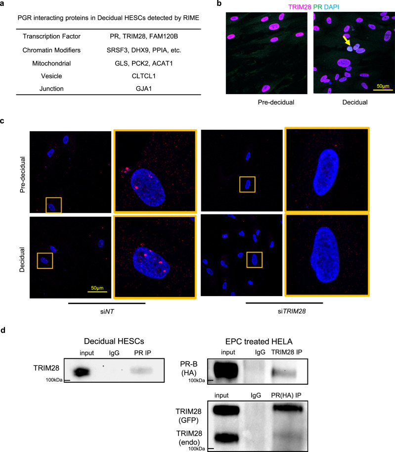
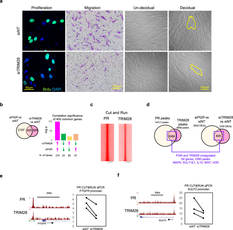
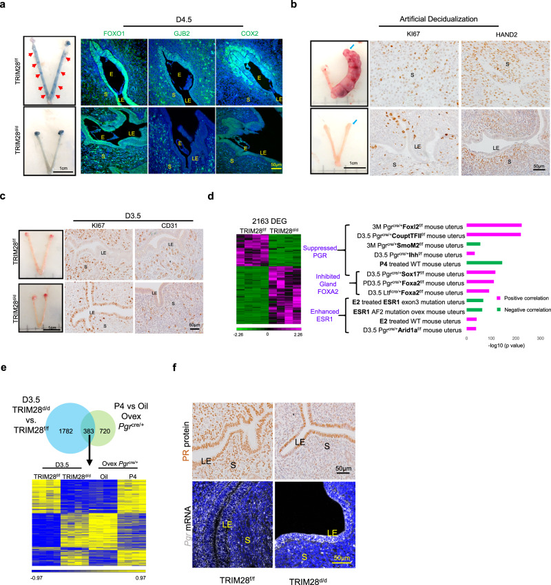
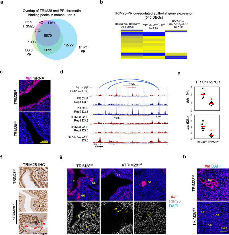
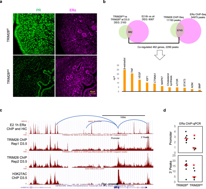
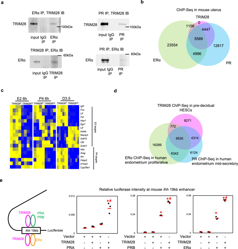
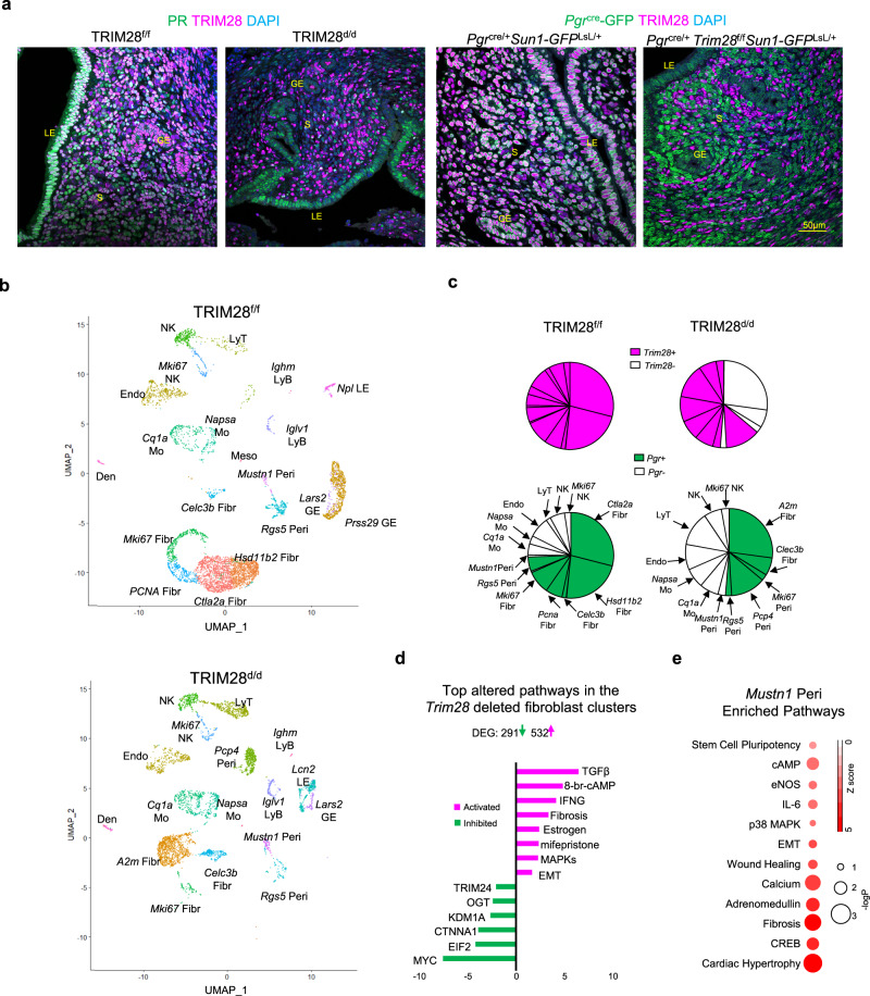
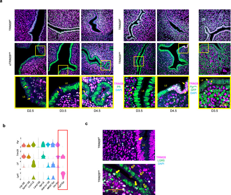
Estrogen and progesterone, acting through their cognate receptors the estrogen receptor α (ERα) and the progesterone receptor (PR) respectively, regulate uterine biology. Using rapid immunoprecipitation and mass spectrometry (RIME) and co-immunoprecipitation, we identified TRIM28 (Tripartite motif containing 28) as a protein which complexes with ERα and PR in the regulation of uterine function. Impairment of TRIM28 expression results in the inability of the uterus to support early pregnancy through altered PR and ERα action in the uterine epithelium and stroma by suppressing PR and ERα chromatin binding. Furthermore, TRIM28 ablation in PR-expressing uterine cells results in the enrichment of a subset of TRIM28 positive and PR negative pericytes and epithelial cells with progenitor potential. In summary, our study reveals the important roles of TRIM28 in regulating endometrial cell composition and function in women, and also implies its critical functions in other hormone regulated systems.


Conflict of interest statement

The authors declare no competing interests.

Figures

Fig. 1
Fig. 1. PGR interacts with TRIM28 to regulate decidualization in HESCs.
a PR interacting proteins in decidual HESCs by RIME. b Immunofluorescence (IF) of TRIM28 (magenta) and PR (green) in pre-decidual and decidual HESCs. c TRIM28 and PR proximity (red dots, arrow pointed) in siNT and siTRIM28 treated pre-decidual and decidual HESCs by Proximity Ligation Assay. d Co-immunoprecipitation of TRIM28 and PR in the decidual HESCs and PR/TRIM28 overexpressed HELA cells. The IF was repeated in three primary cell line. The proximity and co-IP in decidual HESCs were repeated in two primary cell lines. The co-IP in the overexpressed HELA cells was repeated for three independent experiments. All repeats show similar results.
Fig. 2
Fig. 2. TRIM28 knockdown disrupt PR signaling in HESCs.
a Edu labeled cell proliferation, migrated cells in the transwell assay, and brightfield images of pre-decidual and decidual HESCs. Yellow line circles the cells. b Comparison of siPGR and siTRIM28 mediated transcriptomic changes in the hormone stimulated decidual HESCs. c The heatmap of PR and TRIM28 binding peaks in decidual HESCs by CUT&RUN. d The overlapped peaks of TRIM28 and PR, the associated common DEGs altered by TRIM28 and PGR knockdown in the decidual HESCs. Genome browser display of PR and TRIM28 binding peaks, and the CUT&RUN PCR of PR binding activity at PTGFR e and SULF2 f promoter in the siNT or siTRIM28 treated decidual HESCs. N = 500,000 cells examined over three independent experiments and repeated in two primary cell lines with similar results. Two-sided student’s t test. The bight image, cell proliferation, and proliferation were repeated in three primary cell lines with similar results.
Fig. 3
Fig. 3. TRIM28 deletion impaired decidualization and steroid signaling in mouse uterus.
a The representative pictures and immunofluorescence of FOXO1, GJB2, and COX2 at D4.5 uterus. Red arrows pointed to the embryo implantation sites. b The representative uterine pictures and Immunohistochemistry (IHC) of KI67 and HAND2 of decidual uterus upon artificial decidualization. Blue arrow pointed to the decidual horn. c The Immunohistochemistry of proliferative marker KI67 and endothelial marker CD31 at D3.5. d Heatmap and top altered pathways in TRIM28d/d mice at D3.5 based on RNA-seq data. e The comparison of transcriptome from D3.5 TRIM28d/d mice and P4 (progesterone) treated ovariectomized Pgrcre/+mice. f Immunohistochemistry of PR and in situ hybridization of Pgr. E Embryo, LE Luminal epithelium, S Stroma. IHC was repeat in three different mice per group with similar results.
Fig. 4
Fig. 4. TRIM28 deletion suppressed PR signaling at D3.5 uterine epithelium.
a The overlap of TRIM28 and PR ChIP-Seq in the D3.5 mouse uterus, PR in 1 h P4 (progesterone) treated ovariectomized mouse uterus. b The expression heatmap of PR and TRIM28 co-regulated genes. c The in situ hybridization (ISH) of Ihh. d The genome browser of PR, TRIM28 and H3K27AC binding peaks surrounding Ihh. Blue line is the Chromatin loops predicted by HiC. e The ChIP-qPCR of PR at the enhancers 19 kb and 63 kb upstream of Ihh in the TRIM28f/f and TRIM28d/d mice. N = 6 biologically independent samples per group. Two-sided student’s t test. *p < 0.05. f Immunohistochemistry (IHC) of TRIM28 in the D3.5 TRIM28f/f and eTRIM28d/d. g In situ hybridization of Ihh and immunofluorescence of TRIM28 in the D3.5 TRIM28f/f and eTRIM28d/d. h In situ hybridization of Ihh in the D3.5 TRIM28f/f and TRIM28d/d. GE Glandular epithelium. LE luminal epithelium. Rep replicate. ISH and ICH were repeated in three different mice per group with similar results.
Fig. 5
Fig. 5. TRIM28 deletion inhibited ERα activity.
a Immunofluorescence (IF) of PR and ERα. b The overlapped binding peaks of TRIM28 and ERα, the associated common DEGs and top altered pathways between TRIM28d/d mice and E2 treated ovariectomized wildtype mouse uterus. c The genome browser of ERα, TRIM28 and H3K27AC binding peaks at Pgr. Blue line is the Chromatin loops predicted by HiC. d The ChIP-qPCR of ERα binding activity at the promoter and 3′ peak enhancer. Two-sided student’s t test. *p < 0.05. N = 6 biologically independent samples per group. Rep: replicate. The IF was repeated in three different mice per group with similar results.
Fig. 6
Fig. 6. ERα, PR and TRIM28 interacts with each other in the mouse uterus.
a The co-immunoprecipitation (co-IP) of PR, TRIM28, ERα in mouse uterus. IP: immunoprecipitation. IB: Immunoblot. N = 3 mice. b The overlap of TRIM28 ChIP-Seq at D3.5 mouse uterus, PR ChIP-Seq in 1 h Progesterone (P4) treated uterus and ERα ChIP-Seq in 1 h 17β-estradiol (E2) treated uterus. N = 4 mice. c The heatmap of fold changes of E2 and P4 target genes in the P4 6 h or E2 6 h treated ovariectomized TRIM28d/d uterus compared to vehicle, or D3.5 TRIM28d/d uterus compared to the TRIM28f/f uterus. d The overlap of TRIM28 ChIP-Seq in pre-decidual HESCs, and published PR ChIP-Seq in human endometrium at mid-secretory phase, ERα ChIP-Seq in human endometrium at proliferative phase. e Relative luciferase intensity at mouse Ihh 19 kb enhancer in HEC1A cells co-transfected with TRIM28 and PRA or PRB or ERα. N = 150,000 cells examined over three independent experiments. One-way ANOVA with post-hoc turkey’s test. *p < 0.05, compared to vector. #p < 0.05, compared to PRA or PRB or ERα alone. The co-IP was repeated in three different mice with similar results.
Fig. 7
Fig. 7. Abnormally accumulation of TRIM28 positive alone cells in the TRIM28d/d uterus.
a Immunofluorescence (IF) of TRIM28 and PR in the TRIM28f/f and TRIM28d/d mice at D3.5. Immunofluorescence of TRIM28 and Pgrcre-GFP in the Pgrcre/+Sun1-GFPLsL/+ and Pgrcre/+Trim28f/f Sun1-GFPLsL/+ mice at D3.5. b UMAP of scRNA-Seq in the TRIM28f/f and TRIM28d/d mice at D3.5. c The pie plot of all the non-epithelial clusters. Each sector represented one cell cluster. The area of the sector is proportional to the number of the cells in each cluster. The TRIM28+ is magenta. The PGR+ is green. d The top altered pathways in the A2m Fibr from the TRIM28d/d mice compared with the Hsd11b2 Fibr from the TRIM28f/f mice. Green means inhibition. Magenta means activation. e Enriched pathway in Mustn1 Peri annotated by IPA based on the cluster specific marker genes. The size of the red dot is proportional to the -logP value. The red color is proportional to the z score. Fibr Fibroblast, Peri Pericytes, LE Luminal epithelium, GE Glandular epithelium, Endo Endothelium, Mo Myeloid, NK Natural killer cells, LyT Lymphocyte T, LyB Lymphocyte, Den Dendritics, Meso Mesothelium. The IF was repeated in three different mice per group with similar results.
Fig. 8
Fig. 8. Epithelial TRIM28 deletion reduced Ihh expression and promoted progenitor cell accumulations at luminal epithelium.
a The immunofluorescence (IF) of TRIM28 and PR, TRIM28 and Pgrcre-GFP at D2.5, D3.5, and D4.5 of the TRIM28f/f, eTRIM28d/d and TRIM28d/d mouse uterus. Arrow points to the TRIM28 positive cells in the luminal epithelium of the TRIM28d/d and eTRIM28d/d mice. b The violin plot of Pgr, Trim28, Esr1, Lgr5 single cell expressions. c The immunofluorescence of TRIM28 and LGR5. Closed arrow refers to TRIM28 and LGR5 positive epithelium. Open arrow refers to TRIM28 positive but LGR5 negative epithelium. LE Luminal epithelium, GE Glandular epithelium, Epi Epithelium. was repeated in three different mice per group with similar results.
Fig. 9
Fig. 9. TRIM28 signature correlated with human endometrial diseases.
The correlation p value a, c and the T score of gene signature b, d from the D3.5 TRIM28d/d uterus a, b and the siTRIM28 treated decidual HESCs c, d compared with the multiple human endometrial diseases a, c and human endometriosis transcriptome from different subtypes b, d. N = 408 biologically independent samples in total from human endometriosis datasets for b and d. one-way ANOVA with post-hoc turkey’s test was used. *p < 0.05 compared to CE.

References

    1. Azeez JM, et al. New insights into the functions of progesterone receptor (PR) isoforms and progesterone signaling. Am. J. Cancer Res. 2021;11:5214–5232. - PMC - PubMed
    1. Hewitt SC, Korach KS. Estrogen receptors: new directions in the new millennium. Endocr. Rev. 2018;39:664–675. doi: 10.1210/er.2018-00087. - DOI - PMC - PubMed
    1. MacLean JA, 2nd, Hayashi K. Progesterone actions and resistance in gynecological disorders. Cells. 2022;11:647. doi: 10.3390/cells11040647. - DOI - PMC - PubMed
    1. Hewitt, S. C. et al. The estrogen receptor α cistrome in human endometrium and epithelial organoids. bioRxiv 163, bqac116 (2022). - PMC - PubMed
    1. Chi RA, et al. Human endometrial transcriptome and progesterone receptor cistrome reveal important pathways and epithelial regulators. J. Clin. Endocrinol. Metab. 2020;105:e1419–e1439. doi: 10.1210/clinem/dgz117. - DOI - PMC - PubMed

Publication types

MeSH terms