. 2023 Aug 1;21(1):186.
doi: 10.1186/s12964-023-01140-1.

Colonic stem cells from normal tissues adjacent to tumor drive inflammation and fibrosis in colorectal cancer

Affiliations

Colonic stem cells from normal tissues adjacent to tumor drive inflammation and fibrosis in colorectal cancer

Yuanyuan Zhao et al. Cell Commun Signal. .

Abstract

Background: In colorectal cancer (CRC), the normal tissue adjacent to tumor (NAT) communicates actively with the tumor. Adult stem cells from the colon play a crucial role in the development of the colonic epithelium. In the tumor microenvironment, however, it is unclear what changes have occurred in colonic stem cells derived from NAT.

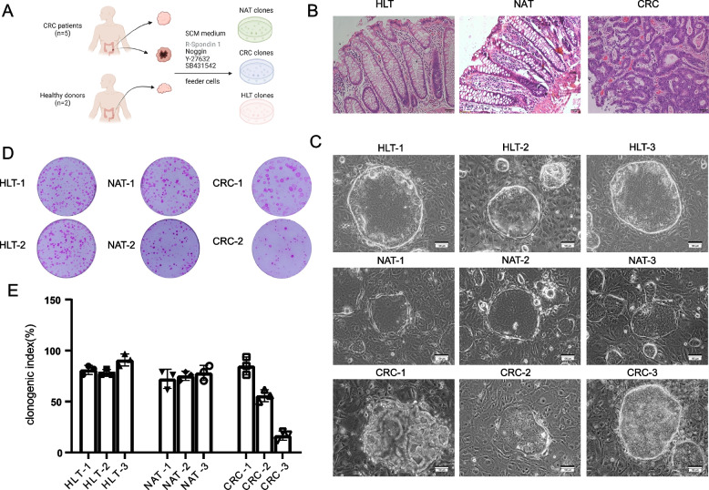
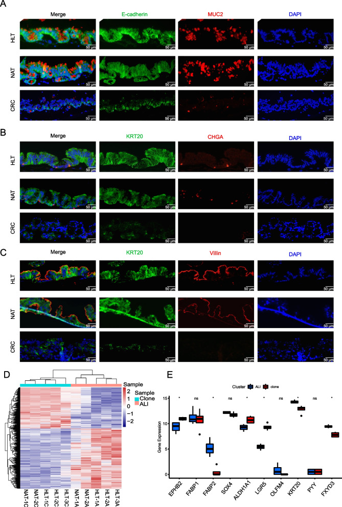
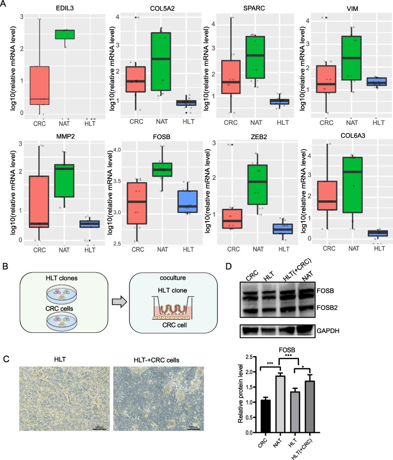
Methods: Using an intestinal stem cell culture system, we cultured colonic cells from NAT and paired CRC tissue, as well as cells from healthy tissue (HLT). Clonogenicity and differentiation ability were used to compare the function of clones from NAT, HLT and CRC tissues. RNA high-throughput sequencing of these clones was used to identify the molecular characteristics of NAT-derived clones. Coculture of clones from HLT and CRC was used to assess molecular changes.

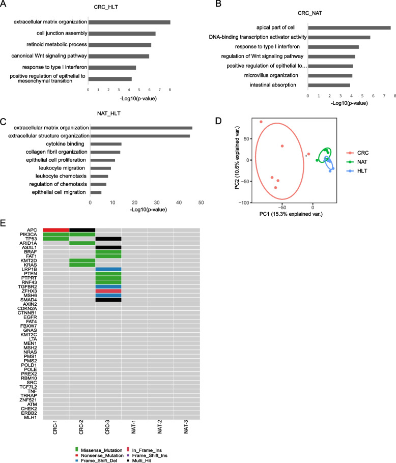
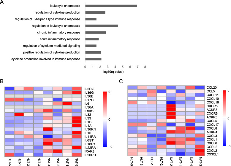
Results: We found that the morphological characteristics, clonogenic ability, and differentiation ability of NAT-derived clones were consistent with those of HLT-derived clones. However, NAT-derived clones changed at the molecular level. A number of genes were specifically activated in NAT. NAT-derived clones enriched pathways related to inflammation and fibrosis, including epithelial mesenchymal transition (EMT) pathway and TGF-beta signaling pathway. Our results also confirmed that NAT-derived clones could recruit fibroblasts in mice. In addition, HLT-derived clones showed high expression of FOSB when cocultured with tumor cells.

Conclusions: Our results demonstrate that colonic stem cells from NAT in the tumor microenvironment undergo changes at the molecular level, and these molecular characteristics can be maintained in vitro, which can induce fibrosis and an inflammatory response. Video Abstract.

Keywords: Colorectal cancer; Fibrosis; Inflammation; Microenvironment; Stem cells.


Conflict of interest statement

The authors declare no competing interests.

Figures

Fig. 1
Fig. 1
Establishment of clones derived from NAT, HLT and tumor tissues. A Overview of experimental design. The clones were derived from NAT and paired CRC tissues, as well as clones from HLT. B Representative H&E staining images of the original tissues. Scale bar, 50 µm. C Representative bright field microscopy images of clones from NAT, HLT and CRC tissues on lawns of irradiated 3T3-J2 feeder cells. Scale bar, 100 µm. D Rhodamine staining showing the clonogenicity assay of colonies grown on feeder cells following seeding 1,000 single cells per well from each clone derived from NAT, HLT and CRC tissues. E Histogram of clonogenicity index based on percentage of plated cells that form colonies. Data represented as mean ± SD; n = 3 technical replicates
Fig. 2
Fig. 2
Differentiation capacity of clones derived from NAT, HLT and tumor tissues. A Immunofluorescence images of ALI mini-guts stained with E-cadherin (green) and Muc2 (red). Nuclei (blue) were stained with DAPI. Scale bar, 50 μm. B Immunofluorescence images of ALI mini-guts stained with KRT20 (green) and CHGA (red). Nuclei (blue) were stained with DAPI. Scale bar, 50 μm. C Immunofluorescence images of ALI mini-guts stained with KRT20 (green) and Villin (red). Nuclei (blue) were stained with DAPI. Scale bar, 50 μm. D Heatmap shows normalized gene expression of stem cell clones (Clone) on feeder cells and corresponding ALI mini-guts (ALI). E Gene expression of stemness markers in stem cells clones and corresponding ALI mini-guts based on RNA-seq data
Fig. 3
Fig. 3
Gene expression differences between clones derived from NAT, HLT and tumor tissues. A Bar plots of the enriched GO terms of upregulated genes in CRC tumor tissues compared with HLT. The terms were selected from the top 20 biological process terms, p < 0.001. B Bar plots of the enriched GO terms of upregulated genes in CRC tumor tissues compared with NAT. The terms were selected from the top 20 biological process terms, p < 0.001. C Bar plots of the enriched GO terms of upregulated genes in NAT compared with HLT. The terms were selected from the top 20 biological process terms. D The principal components analysis (PCA) representing the dispersion of the samples based on their gene expression levels. CRC clones (red), NAT clones (green) and HLT clones (blue). E Somatic mutations found in CRC related genes are present in CRC clones but not in NAT clones. Mutation type is indicated in the legend
Fig. 4
Fig. 4
Inflammatory gene expression by NAT derived clones. A Bar plots of the enriched GO terms of inflammation related genes in clones derived from NAT compared with HLT. B Differential expression heatmaps of interleukin genes among RNA-seq DEGs (FDR < 0.05) of clones derived from NAT and HLT. C Differential expression heatmaps of chemokine genes among RNA-seq DEGs (FDR < 0.05) of clones derived from NAT and HLT
Fig. 5
Fig. 5
NAT derived clones drive myofibroblast activation. A Immunofluorescence micrographs of sections of xenografts formed four weeks after transplantation of clones. Green, E-cadherin; red, Vimentin. Scale bar, 50 μm. B Quantification of fibroblast submucosal accumulation in xenografts based on general scale. Data represented as mean ± SD (Student’s t-test). n = 3 technical replicates. ∗ p < 0.05. C Heatmaps of fibrosis related genes among RNA-seq DEGs (FDR < 0.05) of NAT and HLT. D Gene set enrichment analysis (GSEA) showing the significantly enriched extracellular matrix component pathway (p = 2.06E-4) and EMT pathway (p = 0.0015) in NAT-derived clones
Fig. 6
Fig. 6
HLT-derived clones cocultured with tumor cells. A Genes uniquely expressed in NAT-derived clones compared with HLT and CRC tissues derived clones. B Schematic of HLT-derived clones cocultured with tumor cells using the ALI culture system. C Representative bright field microscopy images of 3D structures formed by HLT-derived clones when cocultured with tumor cells. Scale bar, 100 μm. D Immunoblot analysis showing FOSB expression of HLT-derived clones when cocultured with tumor cells. GAPDH was used as a loading control (left). The band intensity was quantified, data represented the mean ± SD of three independent experiments (right)

References

    1. Siegel RL, Miller KD, Fuchs HE, Jemal A. Cancer statistics, 2022. CA Cancer J Clin. 2022;72(1):7–33. doi: 10.3322/caac.21708. - DOI - PubMed
    1. Zhang L, Li Z, Skrzypczynska KM, Fang Q, Zhang W, O'Brien SA, He Y, Wang L, Zhang Q. Kim A et al: Single-Cell Analyses Inform Mechanisms of Myeloid-Targeted Therapies in Colon Cancer. Cell. 2020;181(2):442–459. doi: 10.1016/j.cell.2020.03.048. - DOI - PubMed
    1. Zhang L, Yu X, Zheng L, Zhang Y, Li Y, Fang Q, Gao R, Kang B, Zhang Q, Huang JY, et al. Lineage tracking reveals dynamic relationships of T cells in colorectal cancer. Nature. 2018;564(7735):268–272. doi: 10.1038/s41586-018-0694-x. - DOI - PubMed
    1. Aran D, Camarda R, Odegaard J, Paik H, Oskotsky B, Krings G, Goga A, Sirota M, Butte AJ. Comprehensive analysis of normal adjacent to tumor transcriptomes. Nat Commun. 2017;8(1):1077. doi: 10.1038/s41467-017-01027-z. - DOI - PMC - PubMed
    1. Trujillo KA, Heaphy CM, Mai M, Vargas KM, Jones AC, Vo P, Butler KS, Joste NE, Bisoffi M, Griffith JK. Markers of fibrosis and epithelial to mesenchymal transition demonstrate field cancerization in histologically normal tissue adjacent to breast tumors. Int J Cancer. 2011;129(6):1310–1321. doi: 10.1002/ijc.25788. - DOI - PMC - PubMed

Publication types