. 2023 Aug 2;19(1):33.
doi: 10.1186/s13005-023-00379-w.

Puricelli biconvex arthroplasty: an experimental study in sheep

Affiliations

Puricelli biconvex arthroplasty: an experimental study in sheep

Renan Langie et al. Head Face Med. .

Abstract

Background: The aim of this study was to establish a sheep model of the Puricelli biconvex arthroplasty (ABiP) technique in sheep for evaluating its functional, biological and histological parameters.

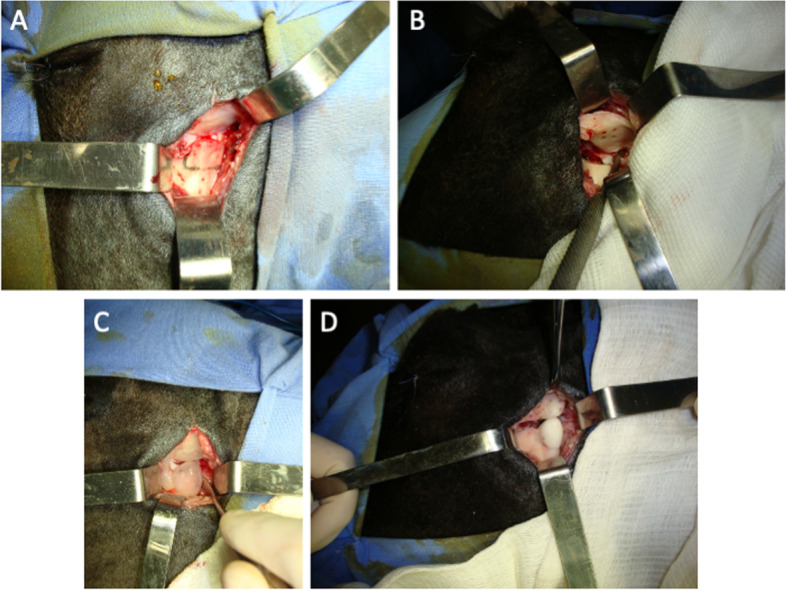
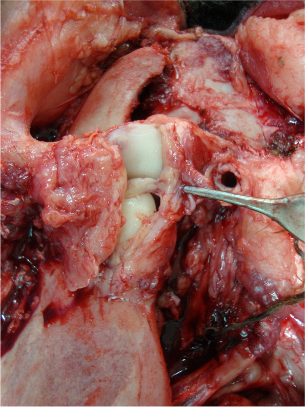
Methods: Ten Corriedale black sheep were submitted to TMJ total reconstruction with poly(methyl methacrylate) (PMMA) using ABiP and euthanized after 45 (n = 5) or 90 (n = 5) days. Control animals (n = 2) underwent sham operations and were euthanized after 45 days. Variables were assessed before the surgery (T0), immediately after (T1) and at 45 or 90 postoperative days (T2).

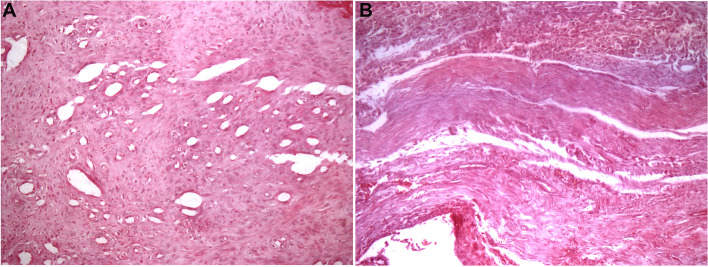
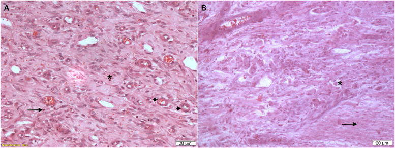
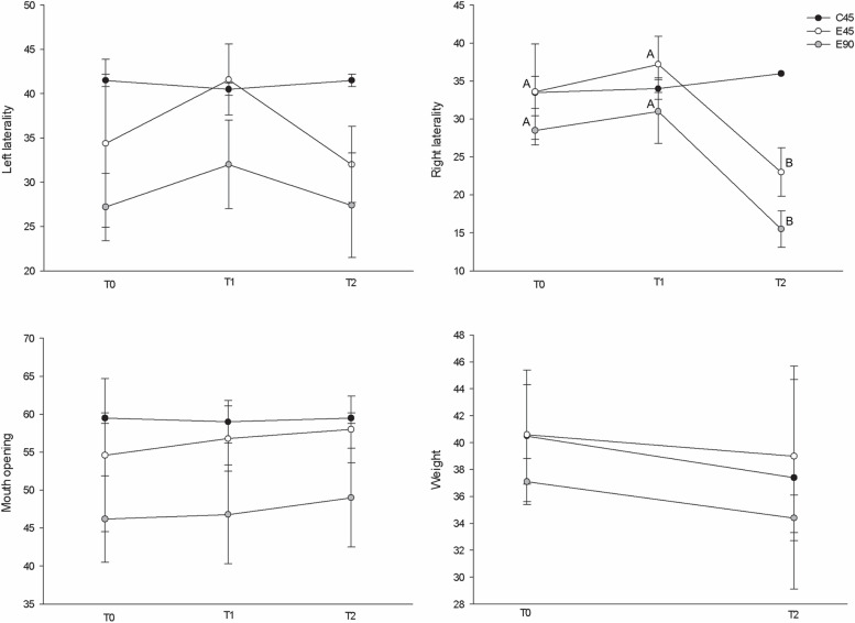
Results: Histological analyses showed regression of inflammatory cells over the follow-up period. PMMA showed reduced porosity and roughness in the articular contact area. PMMA temporal components showed linear and volumetric wear in comparison to control, but no foreign body reaction was observed. The reconstructions were stable in all animals. The amplitude of mouth opening and left lateral movements were maintained, except for a reduction in the range of right lateral movements at day 90 in the experimental group. Clinical, macroscopic and radiographic observations showed that the reconstructions were stable.

Conclusions: The analysis of functional, biological and histological parameters in sheep submitted to ABiP showed stable results of the procedure, with maintenance of body weight and all mandibular movements, save contralateral mandibular movement, suggesting that joint function was completely maintained following the procedure. This experimental study provides support for clinical results previously reported of the ABiP technique in TMJ reconstruction procedures.

Keywords: Arthroplasty; Maxillofacial reconstruction; PMMA; Sheep; Temporomandibular joint; Temporomandibular joint disorders.


Conflict of interest statement

The authors declare no competing interests.

Figures

Fig. 1
Fig. 1
TMJ reconstruction using the Puricelli biconvex arthroplasty technique. A Condylectomy; B Temporal bicortical perforations; C Silicone trays of the temporal and mandibular segments before the reconstruction; D Final reconstruction of the left TMJ
Fig. 2
Fig. 2
Post-mortem TMJ exposure and collection of peri-articular soft tissue samples. The pseudo-disc is macroscopically observed between the temporal and mandibular surfaces of the prosthetic reconstruction
Fig. 3
Fig. 3
Pseudo-disc histological images. A Pseudo-disc at 45 days showing a highly cellularized and vascularized connective tissue (HE, 200x); B Pseudo-disc at 90 days showing a highly fibrous and oriented connective tissue (HE, 200x)
Fig. 4
Fig. 4
Histological capture from the reconstruction capsule showing inflammatory cells at different postoperative times. A 45-day postoperative experimental day. Asterisks, neutrophils; arrows, lymphocytes; arrowheads, angiogenesis; B 90-day postoperative day: asterisks, macrophages; arrows, fibroblasts and collagen fibres. Bar, 20 μm. HE staining
Fig. 5
Fig. 5
Comparison of the degree of conversion (DC) among test and control groups. ImmC (immediate control), 7dC (7-day control), COW (condylar without wear), CWW (condylar with wear), CB (condylar in contact with bone), TOW (temporal without wear), TWW (temporal with wear), TB (temporal in contact with bone). Data are presented as mean and standard deviation, and were compared using an analysis of variance (ANOVA) followed by Tukey’s post hoc tests. Different capital letters represent the presence of statistical differences (p < 0.05)
Fig. 6
Fig. 6
Scanning electron microscopy (100x) – image from PMMA temporal component showing the different pattern of porosity in surface of articular contact. a Surface without wear; b Surface with wear, compatible with the articular contact area
Fig. 7
Fig. 7
Overlapping of the volumetric reconstructions of the temporal (A) and mandibular (B) acrylic components to the corresponding control samples. Red represents a larger volume (mm3) while blue represents a smaller volume (mm3) of the test component in comparison to the control sample
Fig. 8
Fig. 8
Macroscopic and imaging examinations in the postoperative period. A Post-mortem TMJ exposure and collection of peri-articular soft tissue samples. The image shows the clamping of the capsule, which covers the joint reconstruction for the evaluation of reconstruction stability; B Indication of the articular reconstruction site in the X-ray image
Fig. 9
Fig. 9
Comparison between mean maximum mouth-opening amplitudes, left and right lateral movements and animal body weight (in millimetres and kilograms, respectively). For the variable weight the sample size of group E90 was n = 4. Variables are expressed as mean ± standard deviation. Within-group comparisons were performed through repeated-measures analysis of variance (at 3 different moments) or paired-sample T-tests (weight). Letters (A, B) represent significant statistical differences

References

    1. Liu F, Steinkeler A. Epidemiology, diagnosis, and treatment of temporomandibular disorders. Dent Clin North Am. 2013;57(3):465–479. doi: 10.1016/j.cden.2013.04.006. - DOI - PubMed
    1. Onoriobe U, Miloro M. How Many Temporomandibular joint total joint alloplastic implants will be placed in the United States in 2030 ? J Oral Maxillofac Surg. 2016;74(8):1531–1538. doi: 10.1016/j.joms.2016.04.011. - DOI - PubMed
    1. Erdem E, Alkan A. The use of acrylic marbles for interposition arthroplasty in the treatment of temporomandibular joint ankylosis: follow-up of 47 cases. Int J Oral Maxillofac Surg. 2001;30(1):32–36. doi: 10.1054/ijom.2000.0006. - DOI - PubMed
    1. Imola MJ, Liddell A. Temporomandibular joint reconstruction. Curr Opin Otolaryngol Head Neck Surg. 2016;24(4):336–342. doi: 10.1097/MOO.0000000000000282. - DOI - PubMed
    1. Bota NC, Nistor DV, Caterev S, Todor A. Historical overview of hip arthroplasty: from humble beginnings to a high-tech future. Orthop Rev (Pavia) 2021;13(1):8773. doi: 10.4081/or.2021.8773. - DOI - PMC - PubMed

Substances