Review
. 2023 Aug 2;10(1):12.
doi: 10.1186/s44156-023-00024-x.

Three-dimensional printing in modelling mitral valve interventions

Affiliations
Review

Three-dimensional printing in modelling mitral valve interventions

Apurva H Bharucha et al. Echo Res Pract. .

Abstract

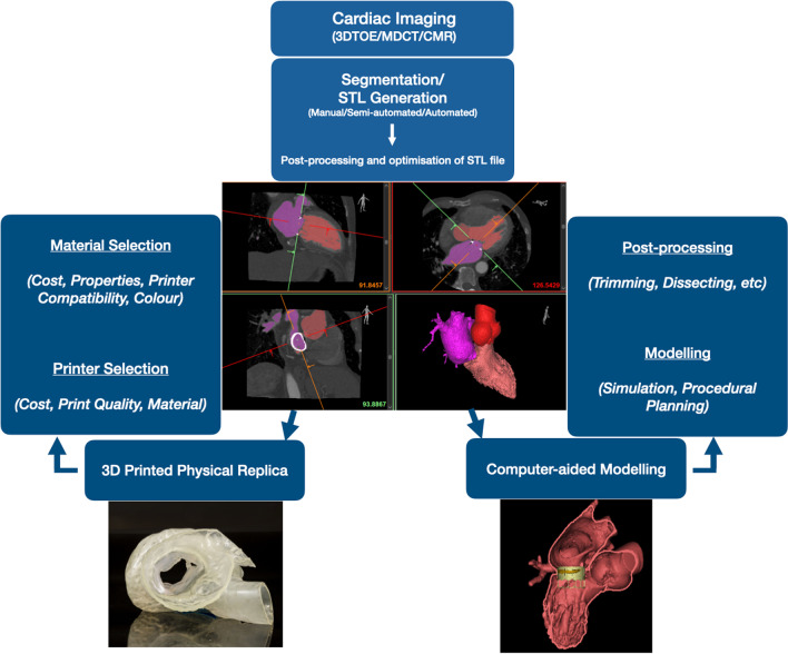
Mitral interventions remain technically challenging owing to the anatomical complexity and heterogeneity of mitral pathologies. As such, multi-disciplinary pre-procedural planning assisted by advanced cardiac imaging is pivotal to successful outcomes. Modern imaging techniques offer accurate 3D renderings of cardiac anatomy; however, users are required to derive a spatial understanding of complex mitral pathologies from a 2D projection thus generating an 'imaging gap' which limits procedural planning. Physical mitral modelling using 3D printing has the potential to bridge this gap and is increasingly being employed in conjunction with other transformative technologies to assess feasibility of intervention, direct prosthesis choice and avoid complications. Such platforms have also shown value in training and patient education. Despite important limitations, the pace of innovation and synergistic integration with other technologies is likely to ensure that 3D printing assumes a central role in the journey towards delivering personalised care for patients undergoing mitral valve interventions.

Keywords: 3D printing; Future technologies; Mitral intervention; Personalised care.


Conflict of interest statement

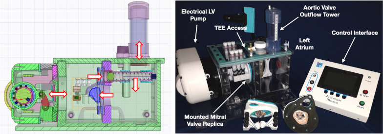
Mr John Moore is a stakeholder in Archetype Biomedical Inc, Ontario, Canada which has manufactured a mitral valve simulator described in the manuscript. The Remainder of the Authors declare no competing interests.

Figures

Fig. 1
Fig. 1
3D printing workflow. 3D printing involves segmentation of a 3D imaging dataset by assigning grey pixels to the anatomical area of interest which is then converted to a file format recognised by a 3D printer. (Most commonly Standard Tessellation Language, STL format). The STL file can be either used for printing out a physical replica or incorporated into virtual platforms for computer-aided design and modelling. 3DTOE Three-Dimensional Echocardiography, MDCT Multi Detector Computational Tomography, CMR Cardiac Magnetic Resonance
Fig. 2
Fig. 2
An overview of 3D printer technologies. A schematic representation of common 3D printer technologies currently available. FDM extrudes thermoplastic filaments layer by layer to generate a 3D replica. SLS uses laser to fuse powder-based polymers. SLA uses a liquid photosensitive resin which coalesces on exposure to laser or UV light irradiation. Polyjet printers extrude a photopolymer which solidifies on exposure to a UV light or laser source and offer the ability to combine multiple polymers to generate physical replicas with complex material properties
Fig. 3
Fig. 3
Application of static patient-specific 3D printing in procedural planning. A 3D printed heart model to assess the risk of LVOTO in patients undergoing TAVI valve in mitral annular calcification procedure. AL anterior mitral leaflet, PL Posterior Mitral leaflet. Adapted from Sabbagh et al. [55]. B Demonstrates the use of a static 3D printed mitral replica for sizing the guide catheter curve within the left ventricular cavity in a patient undergoing direct percutaneous annuloplasty using the Mitralign system. Adapted from Dankowski et al. [52]. C demonstrates an implanted LOTUS TAVI prosthesis within a 3D printed patient-specific mitral replica which was used to aid procedural planning for transapical transcatheter mitral valve implantation. Adapted from Ren et al. [54]
Fig. 4
Fig. 4
3D computer assisted modelling (3DCAM) for transcatheter mitral valve intervention. Patient-specific Multidetector CT data is segmented and the STL file is used to generate a virtual 3D model of patient anatomy which can be evaluated by the Heart Team in multiple planes and used to simulate the trans-catheter mitral valve replacement to assess the neo LVOT and risk of LVOT obstruction (A). The imaging data can also be used to identify optimal fluoroscopic procedural views (B)
Fig. 5
Fig. 5
An example of a Mock Circulatory System (MCS). Left ventricular haemodynamics are simulated by electrical motor which can be tuned by a control interface to modulate physiological variables such as heart rate and stroke volume. Blood mimicking fluid (tap water in this system) is driven through the modelled left heart chambers. A spring within an aortic outflow tower (AV outflow) can be altered to set afterload. A patient-specific silicone valve replica is placed within the system and evaluated using transoesophageal echo. Mitral pathology is simulated by tuning the tension on the chordae of the patient-specific valve replicas [65]
Fig. 6
Fig. 6
Silicone based patient-specific mitral valve replicas using an indirect injection moulding technique. Image A demonstrates a 3D echo dataset used to create a patient-specific rigid mould of the mitral valve in diastole B into which tissue mimicking silicone is applied. Image C shows incorporation of the silicone made mitral valve replica into an assembly that allows integration of the model into the mock circulatory circuit. Valve replicas can be modified per the proposed interventional strategy such as edge-edge mitral leaflet repair D or placement of an annuloplasty ring (E). These replicas can then be mounted and evaluated using transoesaphageal echo within a mock circulatory circuit (F)
Fig. 7
Fig. 7
A model thoracic torso with access ports for  simulating minimally invasive mitral repair (A) housing a silicone cast mitral valve replica (B) mounted within a feedback system assessing suture depth and width (C). Adapted from Sardari Nia et al. [75]
Fig. 8
Fig. 8
Static 3D printed mitral replicas are sufficiently versatile to be integrated with other disruptive technologies such as machine learning and extended realities. Generative adversarial networks (GAN)—a form of machine learning- were used to map patterns learned from intra-operative video sequences in endoscopic mitral valve repair onto the video stream captured during training on non-dynamic mitral surgical simulator using silicone mitral replicas to provide a hyper-realistic training experience. Adapted from Engelhardt et al. [77]

References

    1. Nkomo VT, Gardin JM, Skelton TN, Gottdiener JS, Scott CG, Enriquez-Sarano M. Burden of valvular heart diseases: a population-based study. Lancet. 2006;368(9540):1005–1011. doi: 10.1016/S0140-6736(06)69208-8. - DOI - PubMed
    1. el Sabbagh A, Reddy YNV, Nishimura RA. Mitral valve regurgitation in the contemporary era: insights into diagnosis, management, and future directions. JACC. 2018;11:628–643. - PubMed
    1. Vahanian A, Beyersdorf F, Praz F, Milojevic M, Baldus S, Bauersachs J, et al. ESC/EACTS Guidelines for the management of valvular heart disease. Eur Heart J. 2021 doi: 10.1093/eurheartj/ehab395. - DOI - PubMed
    1. Mirabel M, Iung B, Baron G, Messika-Zeitoun D, Detaint D, Vanoverschelde JL, et al. What are the characteristics of patients with severe, symptomatic, mitral regurgitation who are denied surgery? Eur Heart J. 2007;28(11):1358–1365. doi: 10.1093/eurheartj/ehm001. - DOI - PubMed
    1. Niikura H, Gössl M, Kshettry V, Olson S, Sun B, Askew J, et al. Causes and clinical outcomes of patients who are ineligible for transcatheter mitral valve replacement. JACC. 2019;12(2):196–204. - PubMed