. 2023 Dec;19(12):3221-3229.
doi: 10.1080/15548627.2023.2241125. Epub 2023 Aug 1.

Sustained oral spermidine supplementation rescues functional and structural defects in COL6-deficient myopathic mice

Affiliations

Sustained oral spermidine supplementation rescues functional and structural defects in COL6-deficient myopathic mice

Lisa Gambarotto et al. Autophagy. 2023 Dec.

Abstract

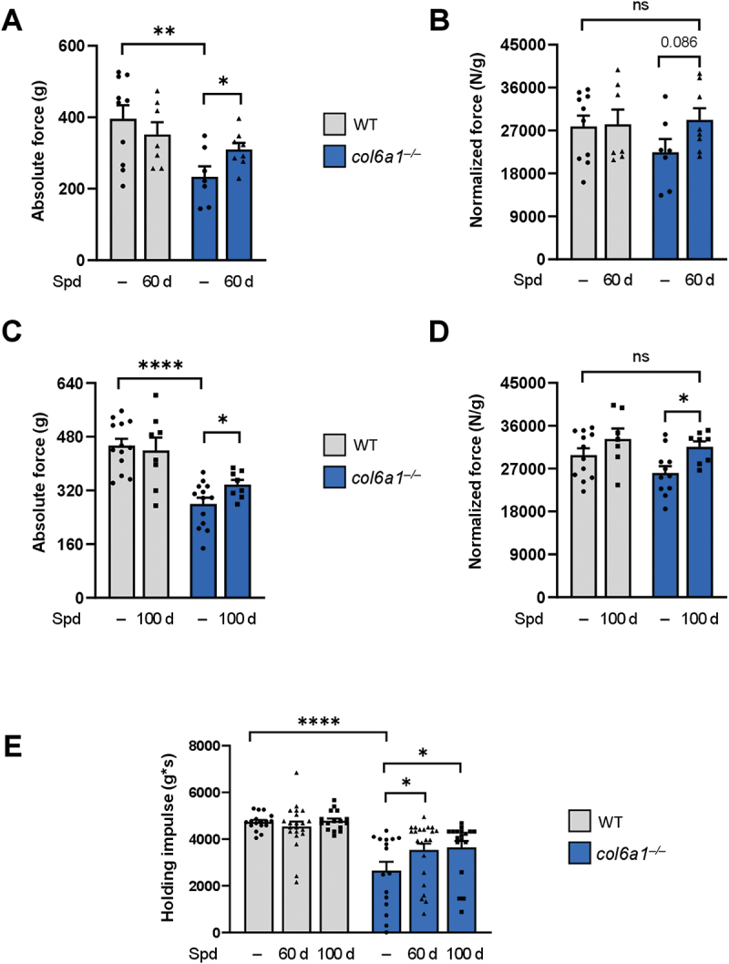
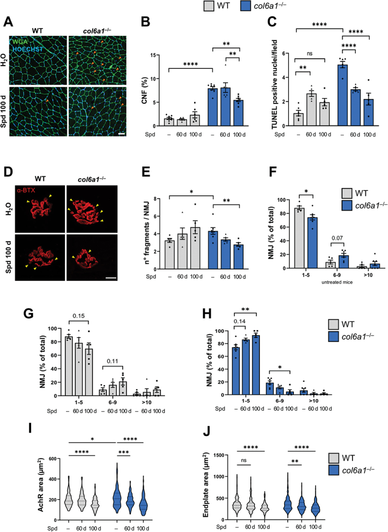
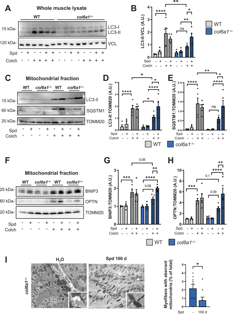
COL6 (collagen type VI)-related myopathies (COL6-RM) are a distinct group of inherited muscle disorders caused by mutations of COL6 genes and characterized by early-onset muscle weakness, for which no cure is available yet. Key pathophysiological features of COL6-deficient muscles involve impaired macroautophagy/autophagy, mitochondrial dysfunction, neuromuscular junction fragmentation and myofiber apoptosis. Targeting autophagy by dietary means elicited beneficial effects in both col6a1 null (col6a1-/-) mice and COL6-RM patients. We previously demonstrated that one-month per os administration of the nutraceutical spermidine reactivates autophagy and ameliorates myofiber defects in col6a1-/- mice but does not elicit functional improvement. Here we show that a 100-day-long spermidine regimen is able to rescue muscle strength in col6a1-/- mice, with also a beneficial impact on mitochondria and neuromuscular junction integrity, without any noticeable side effects. Altogether, these data provide a rationale for the application of spermidine in prospective clinical trials for COL6-RM.Abbreviations: AChR: acetylcholine receptor; BTX: bungarotoxin; CNF: centrally nucleated fibers; Colch: colchicine; COL6: collagen type VI; COL6-RM: COL6-related myopathies; MAP1LC3/LC3: microtubule-associated protein 1 light chain 3; NMJ: neuromuscular junction; Spd: spermidine; SQSTM1/p62: sequestosome 1; TA: tibialis anterior; TOMM20: translocase of outer mitochondrial membrane 20; TUNEL: terminal deoxynucleotidyl transferase dUTP-mediated nick-end labeling.

Keywords: Autophagy; collagen VI; nutraceutical; skeletal muscle; spermidine.


Conflict of interest statement

No potential conflict of interest was reported by the authors.

Figures

Figure 1.
Figure 1.
Long-term Spd treatment rescues muscle strength in col6a1–/– mice. (A-D) In vivo quantification of the absolute tetanic force (panels A and C) and tetanic force normalized on muscle weight (panels B and D) of plantar flexor muscles, at 100 Hz frequency of stimulation, in wild-type and col6a1–/– mice under untreated conditions (–) and after 60 days (60 d, panels A and B) or 100 days (100 d, panels C and D) per os administration of 30 mM Spd. Data are provided as mean±s.e.m. (n = 8–12 muscles, each group; ****, P < 0.0001; **, P < 0.01; *, P < 0.05; ns, not significant; Student’s t-test). (E) Bar plot displaying the hanging impulse, calculated as the recorded hanging time performance normalized per body weight with the four-limb hanging test, in wild-type and col6a1–/– mice under untreated conditions (–) and after 60 days (60 d) or 100 days (100 d) per os administration of 30 mM Spd. Data are provided as mean±s.e.m. (n = 16–22 mice, each group; ****, P < 0.0001; *, P < 0.05; two-way ANOVA test with Holm-Šídák’s post hoc test for multiple comparison). WT, wild type.
Figure 2.
Figure 2.
Long-term Spd treatment ameliorates the histopathological defects of col6a1–/– muscles. (A) Representative images of TA cross-sections from wild-type and col6a1–/– mice treated (Spd 100 d) or not (H2O) with 30 mM Spd administered per os for 100 days. Sections were labeled with wheat germ agglutinin (green) and Hoechst (blue). Orange arrowheads point at centrally nucleated myofibers. Scale bar: 50 µm. (B) Bar plot of the percentage of centrally nucleated myofibers (% CNF), quantified from images as in (A), in wild-type and col6a1–/– mice under untreated conditions (–) and after 60 days (60 d) or 100 days (100 d) per os administration of 30 mM Spd. Data are provided as mean±s.e.m. (n = 6–8, each group; ****, P < 0.00001; **, P < 0.01; two-way ANOVA test with Holm-Šídák’s post hoc test for multiple comparison). (C) Bar plot of the number of TUNEL-positive nuclei in TA cross sections from wild-type and col6a1–/– mice under untreated conditions (–) and after 60 days (60 d) or 100 days (100 d) per os administration of 30 mM Spd. Data are provided as mean±s.e.m. (n = 5–6, each group; ****, P < 0.0001; **, P < 0.01; ns, not significant; two-way ANOVA test with Holm-Šídák’s post hoc test for multiple comparison). (D) Representative confocal max-stack immunofluorescent images of NMJ in diaphragm whole-mount preparations from wild-type and col6a1–/– mice treated (Spd 100 d) or not (H2O) with 30 mM Spd administered per os for 100 days. Sections were labeled with α-bungarotoxin (α-BTX, in red). Yellow arrowheads point at NMJ fragments. Scale bar: 10 µm. (E) Bar plot of the number of AChR fragments per NMJ, quantified from 3D reconstruction of images as in (A), in wild-type and col6a1–/– mice under untreated conditions (–) and after 60 days (60 d) or 100 days (100 d) per os administration of 30 mM Spd. At least 35 NMJ were analyzed for each muscle. Data are provided as mean±s.e.m. (n = 5–7 mice, each group; *, P < 0.05; **, P < 0.01; Mann-Whitney test and Kruskal-Wallis test with Dunn’s post hoc test for multiple comparison). (F-H) Bar plot of NMJ distribution over three separate classes based on the number of AChR fragments (e.g., 1 to 5, 6 to 9, more than 10), quantified from 3D reconstruction of images as in (A), in untreated wild-type and col6a1–/– mice (F), and in wild-type mice (G) and col6a1–/– mice (H) under untreated conditions (–) and after 60 days (60 d) or 100 days (100 d) per os administration of 30 mM Spd treatment. At least 35 NMJ were analyzed for each muscle. Data are provided as mean±s.e.m. (n = 5–7 mice, each group; *, P < 0.05; **, P < 0.01; Mann-Whitney test and Kruskal-Wallis test with Dunn’s post hoc test for multiple comparison). (I-J) Violin plot of AChR area (in µm2, panel I) and of endplate area (in µm2, panel J) of NMJ in wild-type and col6a1–/– mice under untreated conditions (–) and after 60 days (60 d) or 100 days (100 d) per os administration of 30 mM Spd (n = 137–218 NMJ sampled from 5–7 mice, each group; ****, P < 0.0001; ***, P < 0.001; **, P < 0.01; *, P < 0.05; ns, not significant; two-way ANOVA test with Holm-Šídák’s post hoc test for multiple comparison). WT, wild type.
Figure 3.
Figure 3.
Autophagic and mitophagic fluxes of col6a1–/– muscles are reactivated by long-term Spd administration. (A, B) Western blotting (panel A) and densitometric quantification (determined from three independent western blot experiments, panel B) of the non lipidated (LC3-I) and lipidated (LC3-II) forms of LC3B protein in whole muscle protein extracts of wild-type and col6a1–/– mice treated (Spd +) or not (Spd –) with 30 mM Spd administered per os for 100 days, and injected (Colch +) or not (Colch –) with 0.4 mg/kg colchicine. VCL (vinculin) was used as loading control. Data are provided as mean±s.e.m. (n = 4–5 mice, each group; ****, P < 0.0001; **, P < 0.01; *, P < 0.05; ns, not significant; two-way ANOVA test with Holm-Šídák’s post hoc test for multiple comparison). (C-E) Western blotting (panel C) and densitometric quantification (determined from three independent western blot experiments) of the non lipidated (LC3-I) and lipidated (LC3-II) forms of LC3B (panel D) and of SQSTM1/p62 (panel E) in mitochondrial fraction of muscle extracts of wild-type and col6a1–/– mice treated (Spd +) or not (Spd -) with 30 mM Spd administered per os for 100 days, and injected (Colch +) or not (Colch –) with 0.4 mg/kg colchicine. TOMM20 was used as loading control. Data are provided as mean±s.e.m. (n = 4–5 mice, each group; ****, P < 0.0001; **, P < 0.01; *, P < 0.05; two-way ANOVA test with Holm-Šídák’s post hoc test for multiple comparison). (F-H) Western blotting (panel F) and densitometric quantification (determined from three independent western blot experiments) of BNIP3 (panel G) and OPTN1 (panel H) in mitochondrial fraction of muscle extracts of wild-type and col6a1–/– mice treated (Spd +) or not (Spd -) with 30 mM Spd administered per os for 100 days, and injected (Colch +) or not (Colch –) with 0.4 mg/kg colchicine. TOMM20 was used as loading control. Data are provided as mean±s.e.m. (n = 4–5 mice, each group; ****, P < 0.0001; ***, P < 0.001; **, P < 0.01; two-way ANOVA test with Holm-Šídák’s post hoc test for multiple comparison). (I) Representative transmission electron images of diaphragm sections from col6a1–/– mice treated (Spd 100 d) or not (H2O) with 30 mM Spd 30 mM administered per os for 100 days. Bottom squares for each image show higher magnification details of mitochondria. Scale bars: 400 nm. The bar plot on the right shows the quantification of the percentage of myofibers with aberrant mitochondria in col6a1–/– mice in untreated conditions (Spd –) and after 100 days per os administration of 30 mM Spd (100 d). Data are provided as mean±s.e.m. (n = 9, each group; *, P < 0.05; Mann-Whitney test). A.U., arbitrary unit; WT, wild type.

References

    1. Mizushima N. Autophagy: process and function. Genes Dev. 2007;21(22):2861–2873. PubMed PMID: 18006683. doi: 10.1101/gad.1599207 - DOI - PubMed
    1. Dikic I, Elazar Z. Mechanism and medical implications of mammalian autophagy. Nat Rev Mol Cell Biol. 2018;19(6):349–364. PubMed PMID: 29618831. doi: 10.1038/s41580-018-0003-4 - DOI - PubMed
    1. Sandri M. Autophagy in skeletal muscle. FEBS Lett. 2010;584(7):1411–1416. PubMed PMID: 20132819. doi: 10.1016/j.febslet.2010.01.056 - DOI - PubMed
    1. Drake J, Yan Z. Mitophagy in maintaining skeletal muscle mitochondrial proteostasis and metabolic health with ageing. J Physiol. 2017;595(20):6391–6399. PubMed PMID: 28795394. doi: 10.1113/JP274337 - DOI - PMC - PubMed
    1. Bönnemann CG. The collagen VI-related myopathies: Ullrich congenital muscular dystrophy and Bethlem myopathy. Handb Clin Neurol. 2011;101:81–96. PubMed PMID: 21496625. - PMC - PubMed

Publication types