. 2023 Jul 17:11:1168643.
doi: 10.3389/fcell.2023.1168643. eCollection 2023.

Recognition of H2AK119ub plays an important role in RSF1-regulated early Xenopus development

Affiliations

Recognition of H2AK119ub plays an important role in RSF1-regulated early Xenopus development

Saeid Mohammad Parast et al. Front Cell Dev Biol. .

Abstract

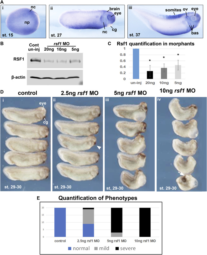
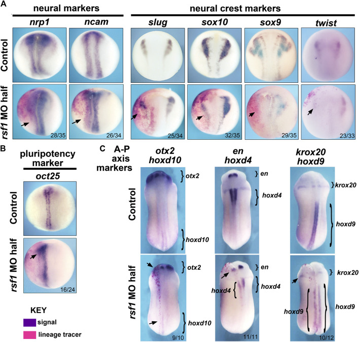
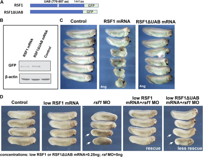
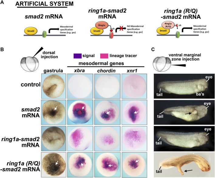
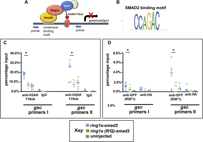
Polycomb group (PcG) proteins are key regulators of gene expression and developmental programs via covalent modification of histones, but the factors that interpret histone modification marks to regulate embryogenesis are less studied. We previously identified Remodeling and Spacing Factor 1 (RSF1) as a reader of histone H2A lysine 119 ubiquitination (H2AK119ub), the histone mark deposited by Polycomb Repressive Complex 1 (PRC1). In the current study, we used Xenopus laevis as a model to investigate how RSF1 affects early embryonic development and whether recognition of H2AK119ub is important for the function of RSF1. We showed that knockdown of Xenopus RSF1, rsf1, not only induced gastrulation defects as reported previously, but specific targeted knockdown in prospective neural precursors induced neural and neural crest defects, with reductions of marker genes. In addition, similar to knockdown of PRC1 components in Xenopus, the anterior-posterior neural patterning was affected in rsf1 knockdown embryos. Binding of H2AK119ub appeared to be crucial for rsf1 function, as a construct with deletion of the UAB domain, which is required for RSF1 to recognize the H2AK119ub nucleosomes, failed to rescue rsf1 morphant embryos and was less effective in interfering with early Xenopus development when ectopically expressed. Furthermore, ectopic deposition of H2AK119ub on the Smad2 target gene gsc using a ring1a-smad2 fusion protein led to ectopic recruitment of RSF1. The fusion protein was inefficient in inducing mesodermal markers in the animal region or a secondary axis when expressed in the ventral tissues. Taken together, our results reveal that rsf1 modulates similar developmental processes in early Xenopus embryos as components of PRC1 do, and that RSF1 acts at least partially through binding to the H2AK119ub mark via the UAB domain during development.

Keywords: H2AK119ub; PRC1; RSF1; UAB domain; Xenopus laevis; development; mesoderm; neural and neural crest.


Conflict of interest statement

The authors declare that the research was conducted in the absence of any commercial or financial relationships that could be construed as a potential conflict of interest.

Figures

FIGURE 1
FIGURE 1
Rsf1 is expressed in multiple tissues of Xenopus laevis during early development and decreasing the levels of Rsf1 causes developmental malformations. (A) Whole mount in situ hybridization of rsf1 mRNA (purple) in Xenopus embryos. Representative lateral views of embryos (anterior to the right) at stages 15, 27 and 37 were shown. Rsf1 sense probe and rsf1 anti-sense probe with no antibody controls were included in Supplementary Figure S1A. (B) Immunoblots showing Rsf1 levels in control and rsf1 MO injected embryos. The amounts of rsf1 MO were indicated on the top of the panel, beta-actin was used as a loading control. Additional and raw images of blots are shown in Supplementary Figure S1C. (C) Quantification of western blots based on three replicates. One-way ANOVA revealed statistical differences among the groups (p < 0.001) and Holm-Sidak posthoc pair-wise comparisons showed statistical differences with each dose and control [p = 0.001 (uninj vs. 5 ng), p = 0.003 (uninj vs. 10 ng), p = 0.006 (ininj vs. 20 ng) SigmaPlot]. (D) Representative images of embryos injected with rsf1 MO revealing malformations that were more severe with increasing concentrations. The amounts of rsf1 MO were indicated on the top of the panel. Anterior is to the right. (E) Quantification of rsf1 morphant embryos displaying various phenotypes. A mild phenotype includes small eyes, reduced head size (e.g., arrowhead). A severe phenotype includes small or no eyes, reduced head size or no head, shortened and bent body axis, failure of blastopore closure (white arrow) (n = 20, 2 biological replicates). nc, neural crest, np, neural plate, cg, cement gland, ov, otic vesicle, bas, branchial arches.
FIGURE 2
FIGURE 2
Targeted knockdown of rsf1 in presumptive neural territories interferes with specification of neural and neural crest tissues and affects anterior-posterior neural patterning. Embryos were all injected with 2.5 ng of MO into one dorsal blastomere. (A) Targeted knockdown of Rsf1 in presumptive neural territories downregulated neural and neural crest marker genes. Dorsal views of representative embryos at stage 17, anterior to the top and the injected side is on the left. The neural (nrp1 and ncam) and neural crest (slug, sox10, sox9, and twist) markers were reduced in the embryos on the side of targeted injection. 100% of controls had expression pattern similar to the representative embryos shown (black arrows, n = 33–35, 2 biological replicates for each marker). (B) The pluripotent marker gene oct25 was expanded on the rsf1 MO injected side. 100% of controls had expression pattern similar to the representative embryos shown (n = 24, 2 biological replicates). (C) Targeted knockdown of rsf1 in presumptive neural territories affected A-P axis gene expression. Dorsal views of representative embryos at stage 24, anterior to the top and the injected side is on the left. Bracketed regions indicate expression domains, and black arrows point to areas where there is a reduced expression. The numbers of the morphant embryos with expression pattern changes similar to those shown are indicated in the bottom right of each panel. 100% of controls had expression pattern similar to the representative embryos shown (n = 10–12, 2 biological replicates).
FIGURE 3
FIGURE 3
The UAB domain is required for RSF1 to regulate Xenopus embryonic development. (A) Schematic representation of the two constructs used in the study consisting of a human RSF fused to GFP and a mutant form of RSF1 with the UAB region deleted and fused to GFP. (B) Immunoblots of RSF protein in extracts prepared from Xenopus embryos injected with RSF1 or RSF1ΔUAB mRNA. The levels of GFP fusion proteins appear the same. β-actin was used as loading controls. Additional images and raw data are included in Supplementary Figure S2A. (C) Embryos injected with 4 ng of RSF1 mRNA resulted in major malformations. However, much less severe developmental defects were observed in embryos injected with 4 ng RSF1ΔUAB mRNA. Representative embryos are shown with anterior to the right (n = 18, two biological replicates). (D) RSF1 but not UAB deleted RSF1ΔUAB rescued the developmental defects caused by rsf1 knockdown. The malformations induced by 5 ng of rsf1 MO were partially rescued by co-injection of low concentrations (0.25 ng) of RSF1 mRNA but not 0.25 ng of RSFΔUAB mRNA (compare embryos with white arrows). Control embryos are un-injected siblings (n = 20, 2 biological replicates). Four representative embryos are shown with anterior to the right.
FIGURE 4
FIGURE 4
Fusion of ring1a to smad2 limits the function of smad2 in mesoderm induction. (A) Schematic showing the experimental system. (B) Embryos dorsally injected with (0.25 ng) of mRNA were examined for changes in pigmentation during gastrulation and inappropriate expression of mesodermal genes, all of which indicate mesoderm induction. Additional pigment and mesodermal gene expression are indicated by white arrows (n = 19–20, two biological replicates). (C) Embryos ventrally injected with (0.25 ng) of mRNA were examined for protrusions, indicatives of secondary axis formation. Overt protrusions are indicated by black arrows. N = 18–20, two biological replicates. ba’s, branchial arches.
FIGURE 5
FIGURE 5
Ring1a-smad2 deposits H2AK119ub marks on the smad2 target gene gsc and recruits RSF1. (A) Schematic of the experimental hypothesis. (B) SMAD2 binding motif. (C) Real-time PCR of input and chromatin immunoprecipitated DNA by the indicated antibody. Embryos were co-injected with GFP-RSF1 (1 ng) and ring1a-smad2 or ring1a-R/Q-smad2 (0.4 ng) and subjected to immunoprecipitation using anti-H2AK119ub antibody. IgG was used as negative controls. ChIP signals were normalized to input signals and percentage inputs were plotted and shown for two primer pairs. (D). Real-time PCR of input and chromatin immunoprecipitated DNA by the indicated antibodies. Embryos were injected as in (C) and subjected to immunoprecipitation using anti-GFP antibody. Anti-HA antibody was used as negative controls. Percentage input results from each technical repeat are represented by symbols and median is shown as a short bar. Wilcoxon matched-pairs signed rank test revealed statistical differences between ring1a-smad2 and ring1a(R/Q)-smad2 groups (p < 0.05). Raw data were included in Supplementary Figure S3B.

Similar articles

Cited by

References

    1. Baile F., Gómez-Zambrano Á., Calonje M. (2022). Roles of Polycomb complexes in regulating gene expression and chromatin structure in plants. Plant Commun. 3, 100267. 10.1016/j.xplc.2021.100267 - DOI - PMC - PubMed
    1. Bajpai R., Chen D. A., Rada-Iglesias A., Zhang J., Xiong Y., Helms J., et al. (2010). CHD7 cooperates with PBAF to control multipotent neural crest formation. Nature 463, 958–962. 10.1038/nature08733 - DOI - PMC - PubMed
    1. Blackledge N. P., Farcas A. M., Kondo T., King H. W., Mcgouran J. F., Hanssen L. L. P., et al. (2014). Variant PRC1 complex-dependent H2A ubiquitylation drives PRC2 recruitment and polycomb domain formation. Cell 157, 1445–1459. 10.1016/j.cell.2014.05.004 - DOI - PMC - PubMed
    1. Blackledge N. P., Klose R. J. (2021). The molecular principles of gene regulation by Polycomb repressive complexes. Nat. Rev. Mol. Cell Biol. 22, 815–833. 10.1038/s41580-021-00398-y - DOI - PMC - PubMed
    1. Bowes J. B., Snyder K. A., Segerdell E., Gibb R., Jarabek C., Noumen E., et al. (2008). Xenbase: A Xenopus biology and genomics resource. Nucleic Acids Res. 36, D761–D767. 10.1093/nar/gkm826 - DOI - PMC - PubMed