Case Reports
. 2023 Aug;64(8):733-741.

Management of obstructive urethroliths, urethral pseudodiverticulum, and stricture by diverticulectomy, urethroplasty, and urethral stenting placement in a male goat

Affiliations
Case Reports

Management of obstructive urethroliths, urethral pseudodiverticulum, and stricture by diverticulectomy, urethroplasty, and urethral stenting placement in a male goat

Florian Wuillemin et al. Can Vet J. 2023 Aug.

Abstract

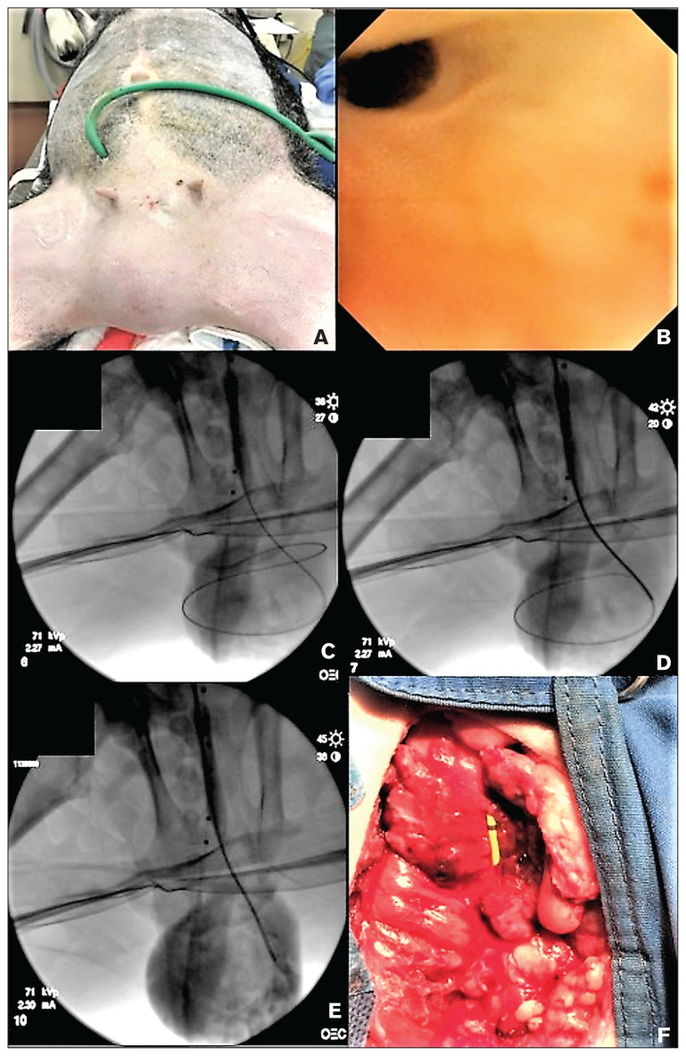
A 5-year-old wether was presented for an acute onset of loss of appetite and inability to urinate. Urethral urolithiasis causing urethral obstruction was diagnosed and a cystostomy catheter was placed. The wether continued to be unable to urinate through the urethra and further developed a perineal pseudodiverticulum. Diverticulectomy followed by a urethroplasty using porcine small intestinal submucosa was performed to relieve the obstruction. The wether developed a urethral stricture following urethroplasty and the owners refused a perineal urethroplasty. Cystourethrography, fluoroscopic-guided balloon dilations, and urethral stent placement were done to establish urethral patency. The wether developed tissue ingrowth through the stent, resulting in recurrent obstruction that necessitated placement of covered urethral stents. Key clinical message: Although obstructive uroliths usually carry a guarded prognosis in small ruminants, the use of novel interventional radiology techniques along with urethroplasty using a xenograft allowed a wether to achieve urethral patency and normal urinations.

Prise en charge d’un bouc présentant des urétrolithes osbtructifs, un pseudodiverticule urétral et une stricture par diverticulectomie, urétroplastie et placement d’un stent urétral. Un bouc castré de 5 ans a été présenté pour une perte aigüe d’appétit et une incapacité à uriner. Un calcul urétral provoquant une obstruction urinaire a été diagnostiquée et une sonde de cystotomie placée. Le bouc a continué d’être incapable d’uriner pas son urètre et a développé un pseudodiverticule périnéal. Une diverticulectomie suivie d’une urétroplastie utilisant de la sous-muqueuse d’intestin grêle de porc a été réalisée pour soulager l’obstruction. Le bouc a développé une stricture urétrale à la suite de l’urétroplastie et les propriétaires ont refusé une urétroplastie périnéale. Une cystourétrographie, des dilatations par ballonnets guidées par fluoroscopie ainsi que le placement d’un stent urétral ont été réalisés afin de résoudre l’obstruction urétrale. Le bouc a développé une réaction tissulaire envahissant la lumière du stent, entrainant ainsi une nouvelle obstruction et nécessitant la mise en place de stents urétraux couverts.Message clinique clé :Bien que les urolithes obstructifs aient généralement un pronostic réservé chez les petits ruminants, l’utilisation de nouvelles procédures provenant de la médecine interventionnelle associées à une urétroplastie utilisant une xénogreffe a permis d’obtenir une perméabilité de son urètre et des mictions normales chez ce bouc.(Traduit par les auteurs).


Figures

Figure 1
Figure 1
Left lateral radiographic view of the goat with a cystostomy catheter. Radiopaque stones are visible in the urethra (black circle) (A). Contrast cystourethrogram performed through the cystostomy catheter shows filling of the urethra up to the stones, which are completely obstructive (B). Left lateral radiographic view following presentation for a firm swelling in the inguinal region. A radiopaque stone along with smaller mineralizations are visible; however, it is unclear if the stones are in the urethra (C). Contrast cystourethrogram shows a pseudodiverticulum communicating with the urethra in the inguinal region, along with urethral obstruction (D). Contrast-enhanced CT showing contrast filling the bladder. The balloon of the Foley cystostomy catheter is visible (E). Contrast-enhanced CT showing fluid-filled diverticulum with 3 attenuating structures (F).
Figure 2
Figure 2
The goat is in dorsal recumbency. A soft-tissue swelling is visible in the inguinal region (A). Antegrade urethroscopy shows a urethral stricture at the site of the pseudodiverticulum (B). Fluoroscopic image showing guidewire access antegrade through the bladder and into the pseudodiverticulum (C). An open-ended catheter is seen advancing over the guidewire (D). The guidewire has been withdrawn and contrast is injected into the pseudodiverticulum (E). The pseudodiverticulum has been debrided and the urethra is being reconstructed. A yellow, open-ended catheter passing through the urethra and bladder helps identify the course of the urethra (F).
Figure 3
Figure 3
A standing voiding cystourethrogram performed by injecting contrast through the cystostomy catheter demonstrates a stricture of the perineal urethra at the site previously reconstructed (A). The goat is anesthetized and in lateral recumbency. A fluoroscopic contrast cystourethrogram carried out by injecting contrast simultaneously antegrade and retrograde allows for measurement of the length of the urethral stricture (B). A urethral dilation balloon is seen passing through the stricture on urethroscopy (C). A urethral balloon passed over a guidewire that has been passed through the bladder and out the penile urethra (through-and-through access) is filled with contrast. Despite maximum pressure, the stricture does not completely efface (D). An uncovered urethral stent is placed so as to completely cover the urethral stricture. Contrast urethrogram shows good urethral patency at the site of the stricture (E). Plain lateral radiograph shows the urethral stent (F).
Figure 4
Figure 4
Cystourethrogram of the goat in right lateral recumbency. Contrast was injected through the cystostomy catheter. A stricture is suspected at the proximal portion of the urethral stent and multiple filling defects are present within the lumen of the stent (A). Filling defects within the lumen of the stent are compatible with tissue ingrowth (B). Cystourethroscopy shows a urethral stricture at the proximal portion of the stent (C). A guidewire passed through the working channel of the scope is visible. Finger-like mucosal proliferations of the urethral mucosa are visible (D). A Holmium:Yag laser fiber passed through the working channel of the scope is visible. The laser was used to section the stricture and make it more amenable to balloon dilation (E). The stricture is partially effaced following lasering and balloon dilation (F). A 2nd covered urethral stent is passed over a guidewire and, with fluoroscopic guidance, is placed within the uncovered urethral stent to restore luminal patency following tissue ingrowth (G). Cystourethroscopy following placement of a covered stent. An open-ended catheter is seen in the lumen and was used to inject contrast to ensure patency following stent placement (H). Urethroscopy showing a covered stent and a T-fastener used to secure the stent in place and prevent migration. A guidewire and catheter can be seen passing through the urethral lumen (I). Standing cystourethrogram showing good urethral patency following placement of a covered stent within an uncovered stent to treat a stricture and obstructive tissue ingrowth. A T-fastener securing the stent to the surrounding tissue to prevent migration is visible (J).
Figure 5
Figure 5
A Mic-Key low-profile gastrostomy tube was placed and used as a cystostomy catheter. The low-profile nature of the tube was appreciated by the goat’s owners and improved the goat’s quality of life as he was no longer required to wear a body band.

References

    1. Leib M, Dinnel H, Ward D, et al. Endoscopic balloon dilation of benign esophageal strictures in dogs and cats. J Vet Intern Med. 2001;15:547–552. - PubMed
    1. Smith MC, Sherman DM. Goat Medicine. 2nd ed. Ames, Iowa: Wiley-Blackwell; 2009. Urinary system; pp. 537–569.
    1. Jones ML, Gibbons PM, Roussel AJ, et al. Mineral composition of uroliths obtained from sheep and goats with obstructive urolithiasis. J Vet Intern Med. 2017;31:1202–1208. - PMC - PubMed
    1. Janke JJ, Osterstock JB, Washburn KE, et al. Use of Walpole’s solution for treatment of goats with urolithiasis: 25 cases (2001–2006) J Am Vet Med Assoc. 2009;234:249–252. - PubMed
    1. Oman RE, Reppert EJ, Streeter RN, et al. Outcome and complications in goats treated by perineal urethrostomy for obstructive urolithiasis: 25 cases (2010–2017) J Vet Intern Med. 2019;33:292–296. - PMC - PubMed

Publication types

LinkOut - more resources