

doi: 10.4103/ijd.ijd_533_22.
Subungual Hyperkeratosis as a Dermoscopic Clue of Primary Fingernail Mycobacterium Marinum Infections
Affiliations
- PMID: 37529440
- PMCID: PMC10389148
- DOI: 10.4103/ijd.ijd_533_22
Item in Clipboard
Subungual Hyperkeratosis as a Dermoscopic Clue of Primary Fingernail Mycobacterium Marinum Infections
Indian J Dermatol.
2023 May-Jun.
No abstract available
Conflict of interest statement
There are no conflicts of interest.
Figures
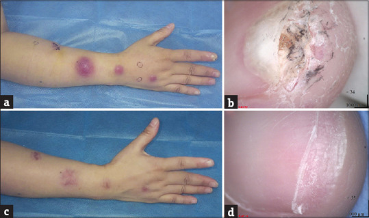
Clinical and dermoscopic findings of the patient infected with Mycobacterium marinum. (a) Multiple disseminated, erythematous, fluctuant nodules distributed in a sporotrichoid pattern on the right upper extremity. (b) Dermoscopic image showing onycholysis, subungual orangish hyperkeratosis surrounded by white scales and fissures of the right index fingernail. (c) After four months, the lesions of the arm were almost healed, leaving residual dark erythema. (d) Advancement of the newly growing nail was observed
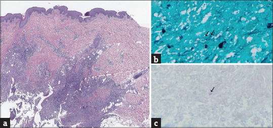
Skin biopsy from the proximal forearm. (a) Chronic suppurative inflammation infiltrated with abscess formation. (b) Gomori methenamine silver staining revealed bacilliform substances (arrow, original magnification ×400). (c) Ziehl–Neelsen staining revealed acid-fast bacilli (arrow, original magnification ×400)
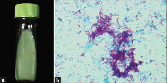
Pus culture and Ziehl–Neelsen staining. (a) Culture showed smooth, cream-yellow colonies (Lowenstein–Jensen media, 30°C, two weeks), which was confirmed as M. marinum by PCR-sequence (GenBank Number: OM649761). (b) Ziehl–Neelsen staining of a colony revealed acid-fast bacilli (original magnification ×1000)
Similar articles
-
Dermoscopic diagnosis of subungual hematoma: new observations.Postepy Dermatol Alergol. 2020 Aug;37(4):490-494. doi: 10.5114/ada.2020.98235. Epub 2020 Sep 2. Postepy Dermatol Alergol. 2020. PMID: 32994768 Free PMC article.
-
A Subungual Exostosis Mimicking a Periungual Granuloma in a 9-Year-Old Boy: An Unexpected Dermoscopic Pattern.Skin Appendage Disord. 2021 Nov;7(6):483-485. doi: 10.1159/000517199. Epub 2021 Jul 9. Skin Appendage Disord. 2021. PMID: 34901181 Free PMC article.
-
Unequal relevance between different subtypes of fingernail psoriasis and psoriatic arthritis.Clin Rheumatol. 2022 Jul;41(7):2065-2069. doi: 10.1007/s10067-022-06107-0. Epub 2022 Feb 19. Clin Rheumatol. 2022. PMID: 35182271
-
[Infections due to Mycobacterium marinum: a review].Hautarzt. 2011 Apr;62(4):266-71. doi: 10.1007/s00105-010-2095-4. Hautarzt. 2011. PMID: 21424893 Review. German.
-
Clinical, dermoscopic, and pathologic features of onychopapilloma: A review of 47 cases.J Am Acad Dermatol. 2016 Mar;74(3):521-6. doi: 10.1016/j.jaad.2015.08.053. Epub 2015 Oct 27. J Am Acad Dermatol. 2016. PMID: 26518173 Review.
References
-
- Bonamonte D, Devito D, Vestita M, Delvecchio S, Ranieri LD, Santantonio M, et al. Aquarium-borne Mycobacterium marinum skin infection. Report of 15 cases and review of the literature. Eur J Dermatol. 2013;23:510–6. - PubMed
-
- Bosamiya SS, Vaishnani JB, Momin AM. Sporotrichoid nocardiosis with cutaneous dissemination. Indian J Dermatol Venereol Leprol. 2011;77:535. - PubMed
-
- Laynez-Roldán P, Fuertes I, Almuedo A, Losada I, Giavedoni P, Camprubí D, et al. Sporotrichoid dissemination of cutaneous leishmaniasis possibly triggered by a diagnostic puncture? J Travel Med. 2020;27:taz044. doi: 10.1093/jtm/taz044. - PubMed
-
- Warren KJ, Fairley JA. Pain and swelling along the nail fold of a 51-year-old man. Dermatol Online J. 1998;4:1. - PubMed
LinkOut - more resources
Full Text Sources