. 2023 Jul 17:14:1215947.
doi: 10.3389/fendo.2023.1215947. eCollection 2023.

Membrane estrogen receptor-α contributes to female protection against high-fat diet-induced metabolic disorders

Affiliations

Membrane estrogen receptor-α contributes to female protection against high-fat diet-induced metabolic disorders

Aurélie Fabre et al. Front Endocrinol (Lausanne). .

Abstract

Background: Estrogen Receptor α (ERα) is a significant modulator of energy balance and lipid/glucose metabolisms. Beyond the classical nuclear actions of the receptor, rapid activation of intracellular signaling pathways is mediated by a sub-fraction of ERα localized to the plasma membrane, known as Membrane Initiated Steroid Signaling (MISS). However, whether membrane ERα is involved in the protective metabolic actions of endogenous estrogens in conditions of nutritional challenge, and thus contributes to sex differences in the susceptibility to metabolic diseases, remains to be clarified.

Methods: Male and female C451A-ERα mice, harboring a point mutation which results in the abolition of membrane localization and MISS-related effects of the receptor, and their wild-type littermates (WT-ERα) were maintained on a normal chow diet (NCD) or fed a high-fat diet (HFD). Body weight gain, body composition and glucose tolerance were monitored. Insulin sensitivity and energy balance regulation were further investigated in HFD-fed female mice.

Results: C451A-ERα genotype had no influence on body weight gain, adipose tissue accumulation and glucose tolerance in NCD-fed mice of both sexes followed up to 7 months of age, nor male mice fed a HFD for 12 weeks. In contrast, compared to WT-ERα littermates, HFD-fed C451A-ERα female mice exhibited: 1) accelerated fat mass accumulation, liver steatosis and impaired glucose tolerance; 2) whole-body insulin resistance, assessed by hyperinsulinemic-euglycemic clamps, and altered insulin-induced signaling in skeletal muscle and liver; 3) significant decrease in energy expenditure associated with histological and functional abnormalities of brown adipose tissue and a defect in thermogenesis regulation in response to cold exposure.

Conclusion: Besides the well-characterized role of ERα nuclear actions, membrane-initiated ERα extra-nuclear signaling contributes to female, but not to male, protection against HFD-induced obesity and associated metabolic disorders in mouse.

Keywords: estrogen receptor alpha (ERα); insulin resistance; membrane-initiated steroid signaling; obesity; sex differences; thermogenesis.


Conflict of interest statement

The authors declare that the research was conducted in the absence of any commercial or financial relationships that could be construed as a potential conflict of interest.

Figures
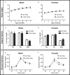
Figure 1
Figure 1
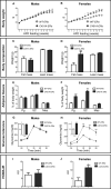
Abrogation of membrane ERα does not influence body weight, adiposity and glucose tolerance in NCD-fed mice. Five-week-old WT-ERα and C451A-ERα male and female mice were maintained on a normal chow diet (NCD) up to 7 months of age. Body weight (A, B), perigonadal (Pg), sub-cutaneous (SC) and mesenteric (Mes) relative fat pad weights (C, D) and intraperitoneal glucose tolerance tests (E and F) are shown in male and female mice, respectively. Data are shown as mean ± SEM (n= 4-14 mice/genotype/sex). (A-D) Unpaired Student’s t test was performed. (E, F) Repeated measures ANOVA were used to compare changes over time between the two genotypes.
Figure 2
Figure 2
C451A-ERα female mice are prone to HFD-induced obesity. Five-week-old WT-ERα and C451A-ERα male and female mice were fed a HFD for 12 weeks. Body weight (A, B), body composition assessed by EchoMRI (C, D), perigonadal (Pg), sub-cutaneous (SC) and mesenteric (Mes) relative fat pad weights (E, F). Intraperitoneal glucose tolerance test (G, H) and HOMA-IR values (I, J) are shown in male and female mice, respectively. AUC: Area under the curve. Data are shown as mean ± SEM (n= 10-15 mice/genotype/sex). (A-F, I, J) Unpaired Student’s t test was performed. (G, H) Repeated measures ANOVA were used to compare changes over time between the two genotypes. For AUC in (G, H), unpaired Student’s t test was performed. *, genotype effect: * p < 0.05, *** p < 0.001.
Figure 3
Figure 3
Abrogation of membrane ERα induces adiposity and liver steatosis in HFD-fed female mice. Five-week-old WT-ERα and C451A-ERα male and female mice were fed a HFD for 12 weeks. Representative pictures (scale bar: 100µm) of perigonadal adipose tissue sections stained with hematoxylin-eosin (H/E) (A), quantification of adipocyte area (B), representative pictures (scale bar: 100µm) of liver histology stained with H/E (top) or oil red O (bottom) (C) and relative quantification of triglycerides and cholesterol esters liver content (D) are shown. Data are shown as mean ± SEM (n= 4-6 mice/genotype). (A-D) Unpaired Student’s t test was performed. *, genotype effect: * p < 0.05, *** p < 0.001.
Figure 4
Figure 4
C451A-ERα female mice are more susceptible to HFD-induced insulin resistance. Five-week-old WT-ERα and C451A-ERα male and female mice were fed a HFD for 12 weeks, then submitted to a hyperinsulinemic-euglycemic clamp procedure. Glucose infusion rate (A) and relative mRNA expression of Glut4 in perigonadal adipose tissue (Pg) and muscle (VL, vastus lateralis) at the end of the clamp (B) are shown. qPCR data were normalized to HPRT and 36B4 mRNA levels for adipose tissue and to Ppia mRNA levels for muscle. In another experimental set, 8-week-old WT-ERα and C451A-ERα female mice were fed a HFD for 6 weeks, then administered with a single insulin intra-peritoneal injection (2U/kg diluted in PBS) or vehicle (PBS alone) after a 12h fasting period and sacrificed 15 minutes later. Western blot analysis of Akt phosphorylation on Ser473 in liver (C) and muscle (VL, vastus lateralis) (D) samples are shown. Gapdh is used as loading controls. Data are shown as mean ± SEM (n= 4-9/genotype). (A-D) Unpaired Student’s t test was performed. *, genotype effect: ** p <0.01, *** p <0.001.
Figure 5
Figure 5
Reduced energy expenditure characterizes HFD-fed C451A-ERα female mice Metabolic parameters were registered in metabolic cages in WT-ERα and C451A-ERα female mice fed a HFD for 4 weeks. Mean values of oxygen consumption (A), CO2 production (B), respiratory quotient (C), energy expenditure (D), physical activity (E) and food intake (F) are shown according to the day (light) and night (dark) periods. Data are shown as mean ± SEM (n= 4/genotype). Two-way ANOVA analyses were used to assess the interaction between the genotype (WT-ERα versus C451A-ERα) and the day period (day versus night). In the absence of significant interaction, statistical analyses regarding the respective influence of genotype and day period are provided. In case of significant interaction, Bonferroni post-tests were subsequently performed to compare the different groups two by two. Genotype effect during the day period: *, p <0.05.
Figure 6
Figure 6
Abolition of membrane ERα signaling alters thermogenesis in HFD-fed C451A-ERα female mice. Five-week-old WT-ERα and C451A-ERα male and female mice were fed a HFD for 12 weeks. Brown adipose tissue (BAT) relative weight (A), representative photomicrograph of BAT sections stained with H/E (scale bar: 100µm) and relative mRNA expression levels of genes involved in thermogenesis and mitochondrial biogenesis (C) are shown. qPCR data were normalized to RPL19 mRNA levels. Another set of HFD-fed female mice were submitted to a 5h cold test (4°C). Evolution of body temperature in response to cold exposure is shown (D). Data are shown as mean ± SEM (n= 4-7/genotype). (A-D) unpaired Student’s t test was performed. *, genotype effect: *p<0.05, **p<0.01.

References

    1. Mauvais-Jarvis F, Clegg DJ, Hevener AL. The role of estrogens in control of energy balance and glucose homeostasis. Endocr Rev (2013) 34:309–38. doi: 10.1210/er.2012-1055 - DOI - PMC - PubMed
    1. Morselli E, Santos RS, Criollo A, Nelson MD, Palmer BF, Clegg DJ. The effects of oestrogens and their receptors on cardiometabolic health. Nat Rev Endocrinol (2017) 13:352–64. doi: 10.1038/nrendo.2017.12 - DOI - PubMed
    1. Tramunt B, Smati S, Grandgeorge N, Lenfant F, Arnal J-F, Montagner A, et al. . Sex differences in metabolic regulation and diabetes susceptibility. Diabetologia (2020) 63:453–61. doi: 10.1007/s00125-019-05040-3 - DOI - PMC - PubMed
    1. Hewitt SC, Korach KS. Estrogen receptors: new directions in the new millennium. Endocr Rev (2018) 39:664–75. doi: 10.1210/er.2018-00087 - DOI - PMC - PubMed
    1. Heine PA, Taylor JA, Iwamoto GA, Lubahn DB, Cooke PS. Increased adipose tissue in male and female estrogen receptor-alpha knockout mice. Proc Natl Acad Sci USA (2000) 97:12729–34. doi: 10.1073/pnas.97.23.12729 - DOI - PMC - PubMed

Publication types

LinkOut - more resources