. 2023 Aug 2;19(8):e1011532.
doi: 10.1371/journal.ppat.1011532. eCollection 2023 Aug.

Broadly neutralizing humanized SARS-CoV-2 antibody binds to a conserved epitope on Spike and provides antiviral protection through inhalation-based delivery in non-human primates

Affiliations

Broadly neutralizing humanized SARS-CoV-2 antibody binds to a conserved epitope on Spike and provides antiviral protection through inhalation-based delivery in non-human primates

Paule Hermet et al. PLoS Pathog. .

Abstract

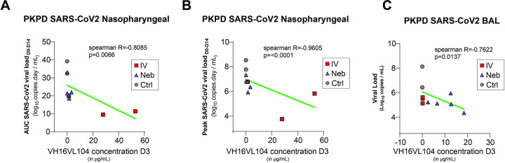
The COVID-19 pandemic represents a global challenge that has impacted and is expected to continue to impact the lives and health of people across the world for the foreseeable future. The rollout of vaccines has provided highly anticipated relief, but effective therapeutics are required to further reduce the risk and severity of infections. Monoclonal antibodies have been shown to be effective as therapeutics for SARS-CoV-2, but as new variants of concern (VoC) continue to emerge, their utility and use have waned due to limited or no efficacy against these variants. Furthermore, cumbersome systemic administration limits easy and broad access to such drugs. As well, concentrations of systemically administered antibodies in the mucosal epithelium, a primary site of initial infection, are dependent on neonatal Fc receptor mediated transport and require high drug concentrations. To reduce the viral load more effectively in the lung, we developed an inhalable formulation of a SARS-CoV-2 neutralizing antibody binding to a conserved epitope on the Spike protein, ensuring pan-neutralizing properties. Administration of this antibody via a vibrating mesh nebulization device retained antibody integrity and resulted in effective distribution of the antibody in the upper and lower respiratory tract of non-human primates (NHP). In comparison with intravenous administration, significantly higher antibody concentrations can be obtained in the lung, resulting in highly effective reduction in viral load post SARS-CoV-2 challenge. This approach may reduce the barriers of access and uptake of antibody therapeutics in real-world clinical settings and provide a more effective blueprint for targeting existing and potentially emerging respiratory tract viruses.


Conflict of interest statement

The authors have declared that no competing interests exist. Patent: 63/318008 Title: Humanized SARS-CoV-2 antibodies Co-inventors: A. Männik, KV, CE, BD, RLG, GK, EJ, PH, MUJ, MU, ES Patent: PCT/IB2021/059363 Title: SARS-Cov-2 neutralizing antibodies Co-inventors: GK, EJ, A.Männik, AP, DK, EŽ, MU, MUJ

Figures

Fig 1
Fig 1. Schematic overview of the HybriFree antibody-cloning procedure and antibody validation workflow.
B-cells from blood of the recovered patient or splenocytes from immunized rabbits were enriched for S-protein antigens and used to develop recombinant antibody expression libraries. Libraries with target specificity were further isolated to single clones followed by biochemical and functional characterization.
Fig 2
Fig 2. Chimeric and humanized variants of rabbit antibody 79C3 show wide neutralization activity against tested SARS-CoV-2 VoCs.
A-F. Both the 79C3 chimeric antibody and ICO-hu104 humanized antibody show neutralizing activity against SARS-CoV-2 VoC Spike protein pseudoviruses in a pseudovirus assay. G. Antibody ICO-hu104 has maintained its pan-neutralizing activity for Omicron sub-variants as tested in a pseudovirus assay.
Fig 3
Fig 3. Antibody ICO-hu104 is detected in the serum and other locations after both intravenous administration and nebulization.
A. Schematic representation of the antibody nebulization system adapted for cynomolgus macaques allowing the localized delivery of antibodies. Rhesus macaque head feature is an asset aquired from BioRender.com. B. Schematic representation of the pharmacology study protocol of non-human primates. Three groups (control [n = 2], intravenous administration [n = 3], and nebulized administration [n = 5]) were exposed to 105 median tissue culture infectious dose (TCID50) of SARS-CoV-2 Delta strain (B.1.617.2) through the intranasal and intratracheal routes. Treated animals received 25 mg/kg of antibody ICO-hu104 one day after the challenge and were sampled over 28 days, as described. For the analysis, we added historical controls challenged under the same conditions (n = 10). C. Antibody ICO-hu104 serum concentrations (mean with SEM). Red represents intravenously administered antibody and blue nebulized antibody. The dotted line represents the LOQ. D. Antibody ICO-hu104 concentration (μg/mL) in heat-inactivated bronchoalveolar lavage (BAL) (left), tracheal fluids (middle), and nasopharyngeal fluids (right) (means ± SD). The dotted line represents the LOQ. C. and D. LOQ = 0,000028 μg/mL, LOD = 0,000009 μg/mL. Red squares represent intravenously administered antibody and blue triangles represent nebulized antibody. Mann-Whitney unpaired two-tailed t-test, *p < 0.05.
Fig 4
Fig 4. Antibody ICO-hu104 retains its integrity and function after nebulization.
A. Analytical size-exclusion chromatography of the ICO-hu104 antibody before and after nebulization. B. Delta and Omicron Spike protein based pseudoviral neutralization assay of ICO-hu104 antibody pre- and post-nebulization.
Fig 5
Fig 5. Antibody ICO-hu104 is capable of reducing the viral load in non-human primates through nebulization and intravenous administration.
A. Genomic (g)RNA in nasopharyngeal fluids is expressed as viral copies per mL. Individual values are plotted by group. Median values are represented by the thick line. The dotted line represents the LOQ = 4760 copies/mL for gRNA (CTRL: control group; in black the extemporaneous controls, IV: intravenous treatment, Neb: Nebulization treatment, LOQ: limit of quantification). B. The area under the curve for data from days 0 to 14 (AUC0-14) of gRNA VL is shown for each individual (Kruskal-Wallis test p<0.0001 followed by Mann-Whitney unpaired two-tailed t-test: **p < 0.01, ***p < 0.001). Black circles represent the 2 extemporaneous control and the grey circles the historical controls, red the squares intravenous treatment group, and blue triangles the nebulization treatment group. C. Subgenomic (sg)RNA determined by RT-qPCR in nasopharyngeal fluids. Individual values are plotted by group. Median values are represented by the thick line. The dotted line represents the LOQ = 7490 copies/mL for sgRNA. Genomic RNA (D) and subgenomic RNA (E) viral loads determined by RT-qPCR of bronchoalveolar lavages (BAL). Individual values are plotted. The bars represent the median values for each group. Kruskal-Wallis test p<0.0001 followed by Mann-Whitney unpaired two-tailed t-test: p** < 0.01, p***< 0.001.
Fig 6
Fig 6. Antibody ICO-hu104 inhibits viral replication.
Viral kinetic parameters (peak and AUC log10 viral load between 0 and 14 dpi) according to the concentration of ICO-hu104 IgG at day 3 in nasopharyngeal fluids with LOD of peak viral load at 2.68. Viral load at day 3 in BAL according to the concentration of ICO-hu104 IgG at day 3 in BAL. A Spearman correlation test was performed to assess the association between Ab concentration and viral kinetic parameters. Two-tailed p value is indicated. Grey: untreated; Red: IV.; Blue: Neb. NS: Nasopharyngeal.
Fig 7
Fig 7. Differential HDX heatmap reveals epitope for ICO-hu23 and ICO-hu104.
Heatmaps (right) show differential HDX uptake at 1, 10, and 60 min. The structures (left) illustrate the location of the epitopes in blue (summed relative fractional uptake difference > 20%) and weaker conformational changes in sky blue (5% A. Wuhan RBD/ICO-hu23. B. Wuhan RBD/ ICO-hu104. C. Omicron RBD/ ICO-hu104.

References

    1. Sabino EC, Buss LF, Carvalho MPS, Prete CA, Crispim MAE, Fraiji NA, et al.. Resurgence of COVID-19 in Manaus, Brazil, despite high seroprevalence. The Lancet. 2021;397(10273):452–5. doi: 10.1016/S0140-6736(21)00183-5 - DOI - PMC - PubMed
    1. Graham MS, Sudre CH, May A, Antonelli M, Murray B, Varsavsky T, et al.. The effect of SARS-CoV-2 variant B.1.1.7 on symptomatology, re-infection and transmissibility. medRxiv. 2021:2021.01.28. doi: 10.1101/2021.01.28.21250680 - DOI
    1. Volz E, Mishra S, Chand M, Barrett JC, Johnson R, Geidelberg L, et al.. Transmission of SARS-CoV-2 Lineage B.1.1.7 in England: Insights from linking epidemiological and genetic data. medRxiv. 2021:2020.12.30. doi: 10.1101/2020.12.30.20249034 - DOI
    1. Chen P, Nirula A, Heller B, Gottlieb RL, Boscia J, Morris J, et al.. SARS-CoV-2 Neutralizing Antibody LY-CoV555 in Outpatients with Covid-19. N Engl J Med. 2021;384(3):229–37. Epub 20201028. doi: 10.1056/NEJMoa2029849 ; PubMed Central PMCID: PMC7646625. - DOI - PMC - PubMed
    1. Weinreich DM, Sivapalasingam S, Norton T, Ali S, Gao H, Bhore R, et al.. REGN-COV2, a Neutralizing Antibody Cocktail, in Outpatients with Covid-19. N Engl J Med. 2021;384(3):238–51. Epub 20201217. doi: 10.1056/NEJMoa2035002 ; PubMed Central PMCID: PMC7781102. - DOI - PMC - PubMed

Publication types

Grants and funding

LinkOut - more resources