. 2023 Aug 2;9(31):eadg6856.
doi: 10.1126/sciadv.adg6856. Epub 2023 Aug 2.

CCR2 is a host entry receptor for severe fever with thrombocytopenia syndrome virus

Affiliations

CCR2 is a host entry receptor for severe fever with thrombocytopenia syndrome virus

Leike Zhang et al. Sci Adv. .

Abstract

Severe fever with thrombocytopenia syndrome virus (SFTSV) is an emerging tick-borne bunyavirus causing a high fatality rate of up to 30%. To date, the receptor mediating SFTSV entry remained uncharacterized, hindering the understanding of disease pathogenesis. Here, C-C motif chemokine receptor 2 (CCR2) was identified as a host receptor for SFTSV based on a genome-wide CRISPR-Cas9 screen. Knockout of CCR2 substantially reduced viral binding and infection. CCR2 enhanced SFTSV binding through direct binding to SFTSV glycoprotein N (Gn), which is mediated by its N-terminal extracellular domain. Depletion of CCR2 in C57BL/6J mouse model attenuated SFTSV replication and pathogenesis. The peripheral blood primary monocytes from elderly individuals or subjects with underlying diabetes mellitus showed higher CCR2 surface expression and supported stronger binding and replication of SFTSV. Together, these data indicate that CCR2 is a host entry receptor for SFTSV infection and a novel target for developing anti-SFTSV therapeutics.


Figures

Fig. 1.
Fig. 1.. CCR2 is required for efficient SFTSV infection in cells.
(A to C) RT-qPCR (A), flow cytometry (B), and Western blot (C) analysis of SFTSV infection at 24 hours after infection in CCR2- or ATF6-knockdown THP-1 cells. n = 6. GAPDH, glyceraldehyde-3-phosphate dehydrogenase. (D) RT-qPCR analysis of virus RNA in CCR2-KO THP-1 cells inoculated with four phylogenetically distinct SFTSV strains, including HBMC16, HNXY2017-50, HNXY2017-66, and WCH, for 24 hours. n = 4. (E) Multistep growth curves of four SFTSV strains in CCR2-KO THP-1 cells. n = 4. (F) Surface expression of CCR2 on BMDMs from CCR2−/− and WT C57BL/6J mice. A representative of three replicates is shown. (G and H) Statistical results (G) and scanned images (H) of the immunological focus assay of SFTSV titers at 24 hours after infection in BMDMs with deletions in CCR2. n = 6. (I) Microscopy of BMDMs immunostained for F4/80, SFTSV NP, and DAPI at 24 hours after infection. (J) SFTSV infection rates at 24 hours after infection determined by flow cytometry analysis in Huh7, HeLa, and Jurkat cells overexpressing CCR2A or CCR2B. n = 6. (K) Representative flow plot of SFTSV infection in Huh7 cells. (L) Supernatant viral titers measured by immunological focus assay in Huh7, HeLa, and Jurkat cells overexpressing CCR2A or CCR2B. n = 6. Two-tailed Student’s t test was performed for comparison of variables between two groups [(A), (B), (D), and (G)]. One-way ANOVA followed by Tukey’s multiple comparisons test was performed for comparison of variables among three groups [(J) and (K)].
Fig. 2.
Fig. 2.. Effect of CCR2 inhibitor and antibody on SFTSV infection in cells.
(A to F) Effects of CCR2 antagonist RS102895 and CCR2 antagonist 1 on SFTSV infection in THP-1 (A) to (C) and Huh7 cells (D) to (F). At 24 hours after infection, relative vRNA levels were measured via RT-qPCR [(A), (B), (D), and (E), n = 3], and relative intracellular SFTSV NP levels were measured by Western blotting (C) and (F). Cell viability was measured using CCK-8 [(D) and (E), n = 3]. (G to I) Effects of CCR2 antibody on SFTSV infection in THP-1 cells. The dose-dependent inhibitory effects of the CCR2 antibody were analyzed by detecting virus loads in THP-1 cells at 24 hours after infection [(G), n = 6]. SFTSV infection rates were measured at 24 hours after infection with MOIs of 1 and 5 [(H), n = 6]. Representative flow plot of SFTSV infection in THP-1 cells treated with anti-human CCR2 or isotype control antibody (I). Two-tailed Student’s t test was performed for comparison of variables between two groups [(A), (B), (D), (E), (G), and (H)]. R2 [(A), (B), (D), and (E)] was estimated by a nonlinear regression model (curve fit).
Fig. 3.
Fig. 3.. CCR2 mediates SFTSV binding and internalization.
(A and B) Effects of CCR2 deletions on the internalization (A) and binding (B) of SFTSV in BMDMs. Relative vRNA levels were measured via RT-qPCR. n = 6. (C) Fluorescence microscopy analysis of the effects of CCR2 deletions on the binding of SFTSV. BMDMs were immunostained with F4/80 (green), SFTSV NP (red), and DAPI. A representative of three replicates is shown. (D and E) SFTSV binding assay in CCR2-KO (KO) THP-1 cells by using RT-qPCR (D) and immunofluorescence staining (E). n = 4. Cellular membranes were labeled with WGA. (F) Flow cytometry analysis of SFTSV infection at 2 hours after infection in Huh7, HeLa, and Jurkat cells overexpressing CCR2A or CCR2B. n = 6. (G to I) Effects of the CCR2 antagonist RS102895 (G), CCR2 antagonist 1 (H), and favipiravir (I) on the binding and internalization of SFTSV in THP-1 cells. n = 3. (J) Binding assay of HRTV, RVFV, and AMRV for control and CCR2-KO THP-1 cells. n = 3. (K and L) Effects of CCR2 antagonist RS102895 (K) and CCR2 antagonist 1 (L) on the binding of HRTV, RVFV, and AMRV in THP-1 cells. n = 3. Two-tailed Student’s t test was performed for comparison of variables between two groups [(A), (B), (D), and (J)]. One-way ANOVA followed by Tukey’s multiple comparisons test was performed for comparison of variables among three groups [(F) to (I), (K), and (L)]. ns, no significance; p.i., post-infection.
Fig. 4.
Fig. 4.. The CCR2 N-terminal extracellular domain mediates SFTSV binding to cells.
(A) Coimmunoprecipitation of co-overexpressed Gn-strep protein and CCR2-flag proteins in HEK293T cells using strep-tag binding beads or anti-Flag antibody beads. Vector-flag, GFP-flag, and SCARB1-flag proteins were used as negative controls. (B to D) Effect of CCR2 overexpression on the infectivity of SFTSV in Huh7 (B), HeLa (C), and Jurkat (D) cells. SFTSV infection rates were measured at 24 hours after infection by flow cytometry analysis in control-, CCR2A-, CCR2B-, CCR2A-ΔN–, and CCR2B-ΔN–overexpressing cells. N = 6. (E) Surface expression of CCR2 and representative flow plot of SFTSV infection in Jurkat cells. (F and G) Effect of the CCR2 N-terminal extracellular domain on the infectivity of SFTSV in HeLa (F) and Jurkat (G) cells. SFTSV infection rates were measured at 24 hours after infection by flow cytometry in control-, CCR2A-, CCR2A-N14Q–, CCR2A-Y26F–, CCR2B-, CCR2B-N14Q–, and CCR2B-Y26F–overexpressing cells. n = 6. (H) Binding of CCR2 N-terminal–derived Y26 sulfated peptide (p2), peptide without tyrosine sulfation at Y26 (p1), or the scrambled peptide to SFTSV virions determined by PRM assay. Statistical analysis is shown in the left panel, and PRM transitions are shown in the right panel. (I and J) Effect of CCR2 N-terminal–derived peptides on the infectivity of SFTSV in THP-1 cells. Relative intracellular vRNA levels and supernatant viral titers were measured at 24 hours after infection via RT-qPCR. n = 4. (K) Effect of CCR2 N-terminal–derived peptides on the binding of SFTSV in THP-1 cells. Relative levels of bound virions were measured via RT-qPCR. n = 4. One-way ANOVA followed by Tukey’s multiple comparisons test was performed for comparison of variables among three groups [(B) to (D) and (F) to (K)].
Fig. 5.
Fig. 5.. CCR2 contributes to SFTSV pathogenesis in mouse models.
(A) Serum and spleen viral titers in SFTSV-infected CCR2−/− and WT C57BL/6J mice (four for each group) tested by immunological focus assay at 3 and 5 dpi. (B and C) Survival probability (B) and relative body weight (C) in anti-IFNAR1 antibody–pretreated CCR2−/− (n = 11) and WT (n = 11) C57BL/6J mice after intraperitoneal infection with SFTSV. (D) Viral titers in serum, spleen, liver, and lung samples from anti-IFNAR1 antibody–pretreated CCR2−/− and WT C57BL/6J mice (four for each group) tested by immunological focus assay at 3 and 5 dpi. (E and F) Representative images of spleen, liver, and lung sections collected at 5 dpi from control and SFTSV-challenged CCR2−/− and WT C57BL/6J mice stained with a rabbit polyclonal antibody against SFTSV NP (E) or with hematoxylin and eosin (F). (G) Viral titers in serum from anti-IFNAR1 antibody–pretreated C57BL/6J mice tested by immunological focus assay at 3 (n = 13 for each group) and 5 (n = 10 for nontreated group and n = 13 for treated group) dpi. (H) Survival probability in anti-IFNAR1 antibody–pretreated C57BL/6J mice with SFTSV infection in the absence (n = 13) or presence (n = 13) of CCR2 antagonist RS102895 and without SFTSV infection (n = 5). Two-tailed Student’s t test was performed for comparison of variables between two groups [(A), (B), (D), and (G)]. The Kaplan-Meier method was used to analyze time-to-event data [(C) and (H)].
Fig. 6.
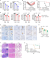
Fig. 6.. CCR2 contributes to SFTSV infectivity in primary human monocytes.
(A and B) Comparisons of surface CCR2 expression levels on primary human monocytes (left) and the ability of SFTS binding (right) between donors aged <60 (n = 10) and ≥60 (n = 10) years old (A), as well as between healthy donors (n = 8) and donors with DM (n = 8) (B). MFI, mean fluorescent intensity. (C) Association of surface CCR2 expression level on primary human monocytes with virus binding ability or with the age of donors (n = 20). Ra2 indicates the correlation between the CCR2 expression level and individual age, and Rb2 indicates the correlation between the CCR2 expression level and the binding ability of SFTSV. (D) Association of surface CCR2 expression level on primary human monocytes with the peak viral load in serum that was consecutively collected from SFTS patients (n = 45) during the clinical course. Two-tailed Student’s t test was performed for comparison of variables between two groups [(A) and (B)]. R2 [(C) and (D)] was estimated by a linear regression model. HC, healthy control; DM, diabetes mellitus; CT, cycle threshold.

References

    1. V. S. Balakrishnan, WHO launches global initiative for arboviral diseases. Lancet Microbe 3, e407 (2022). - PMC - PubMed
    1. X. J. Yu, M. F. Liang, S. Y. Zhang, Y. Liu, J. D. Li, Y. L. Sun, L. Zhang, Q. F. Zhang, V. L. Popov, C. Li, J. Qu, Q. Li, Y. P. Zhang, R. Hai, W. Wu, Q. Wang, F. X. Zhan, X. J. Wang, B. Kan, S. W. Wang, K. L. Wan, H. Q. Jing, J. X. Lu, W. W. Yin, H. Zhou, X. H. Guan, J. F. Liu, Z. Q. Bi, G. H. Liu, J. Ren, H. Wang, Z. Zhao, J. D. Song, J. R. He, T. Wan, J. S. Zhang, X. P. Fu, L. N. Sun, X. P. Dong, Z. J. Feng, W. Z. Yang, T. Hong, Y. Zhang, D. H. Walker, Y. Wang, D. X. Li, Fever with thrombocytopenia associated with a novel bunyavirus in China. N. Engl. J. Med. 364, 1523–1532 (2011). - PMC - PubMed
    1. Y. R. Kim, Y. Yun, S. G. Bae, D. Park, S. Kim, J. M. Lee, N. H. Cho, Y. S. Kim, K. H. Lee, Severe fever with thrombocytopenia syndrome virus infection, South Korea, 2010. Emerg. Infect. Dis. 24, 2103–2105 (2018). - PMC - PubMed
    1. T. Takahashi, K. Maeda, T. Suzuki, A. Ishido, T. Shigeoka, T. Tominaga, T. Kamei, M. Honda, D. Ninomiya, T. Sakai, T. Senba, S. Kaneyuki, S. Sakaguchi, A. Satoh, T. Hosokawa, Y. Kawabe, S. Kurihara, K. Izumikawa, S. Kohno, T. Azuma, K. Suemori, M. Yasukawa, T. Mizutani, T. Omatsu, Y. Katayama, M. Miyahara, M. Ijuin, K. Doi, M. Okuda, K. Umeki, T. Saito, K. Fukushima, K. Nakajima, T. Yoshikawa, H. Tani, S. Fukushi, A. Fukuma, M. Ogata, M. Shimojima, N. Nakajima, N. Nagata, H. Katano, H. Fukumoto, Y. Sato, H. Hasegawa, T. Yamagishi, K. Oishi, I. Kurane, S. Morikawa, M. Saijo, The first identification and retrospective study of severe fever with thrombocytopenia syndrome in Japan. J. Infect. Dis. 209, 816–827 (2014). - PMC - PubMed
    1. H. Li, Q. B. Lu, B. Xing, S. F. Zhang, K. Liu, J. Du, X. K. Li, N. Cui, Z. D. Yang, L. Y. Wang, J. G. Hu, W. C. Cao, W. Liu, Epidemiological and clinical features of laboratory-diagnosed severe fever with thrombocytopenia syndrome in China, 2011–17: A prospective observational study. Lancet Infect. Dis. 18, 1127–1137 (2018). - PubMed