

Neutral ceramidase-active site inhibitor chemotypes and binding modes
- PMID: 37531819
- PMCID: PMC10681040
- DOI: 10.1016/j.bioorg.2023.106747
Neutral ceramidase-active site inhibitor chemotypes and binding modes
Abstract
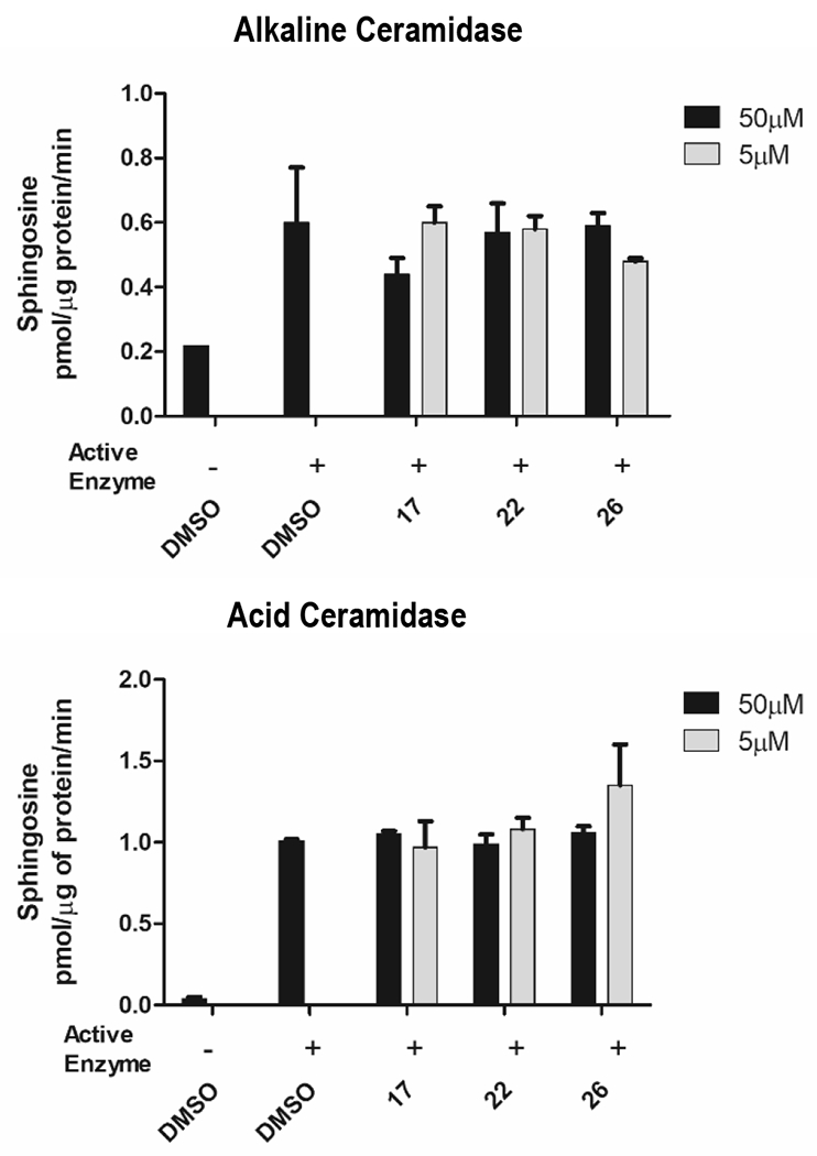
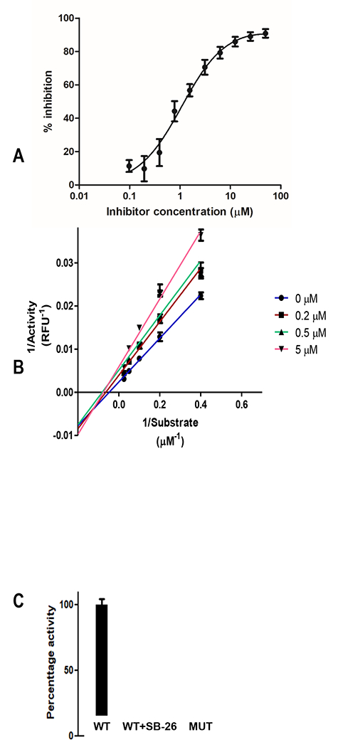
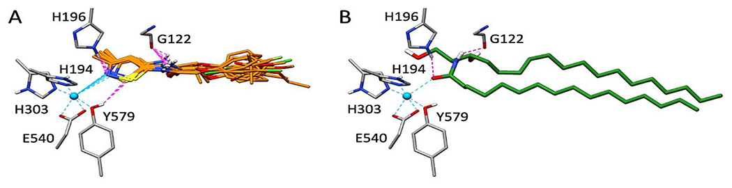
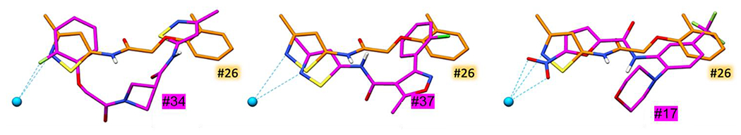
Ceramides impact a diverse array of biological functions and have been implicated in disease pathogenesis. The enzyme neutral ceramidase (nCDase) is a zinc-containing hydrolase and mediates the metabolism of ceramide to sphingosine (Sph), both in cells and in the intestinal lumen. nCDase inhibitors based on substrate mimetics, for example C6-urea ceramide, have limited potency, aqueous solubility, and micelle-free fraction. To identify non-ceramide mimetic nCDase inhibitors, hit compounds from an HTS campaign were evaluated in biochemical, cell based and in silico modeling approaches. A majority of small molecule nCDase inhibitors contained pharmacophores capable of zinc interaction but retained specificity for nCDase over zinc-containing acid and alkaline ceramidases, as well as matrix metalloprotease-3 and histone deacetylase-1. nCDase inhibitors were refined by SAR, were shown to be substrate competitive and were active in cellular assays. nCDase inhibitor compounds were modeled by in silico DOCK screening and by molecular simulation. Modeling data supports zinc interaction and a similar compound binding pose with ceramide. nCDase inhibitors were identified with notably improved activity and solubility in comparison with the reference lipid-mimetic C6-urea ceramide.
Keywords: Colorectal cancer; Neutral ceramidase; Neutral ceramidase inhibitor; nCDase.
Copyright © 2023 Elsevier Inc. All rights reserved.
Conflict of interest statement
Declaration of Competing Interest The authors declare that they have no known competing financial interests or personal relationships that could have appeared to influence the work reported in this paper.
Figures
References
-
- Duan RD, Nilsson A, Metabolism of sphingolipids in the gut and its relation to inflammation and cancer development, Prog Lipid Res 48(1) (2009) 62–72. - PubMed
-
- Hannun YA, Obeid LM, Principles of bioactive lipid signalling: lessons from sphingolipids, Nature reviews Molecular cell biology 9(2) (2008) 139–150. - PubMed
-
- Morad SA, Cabot MC, Ceramide-orchestrated signalling in cancer cells, Nature Reviews Cancer 13(1) (2012) 51–65. - PubMed
-
- Ogretmen B, Hannun YA, Biologically active sphingolipids in cancer pathogenesis and treatment, Nature Reviews Cancer 4(8) (2004) 604–616. - PubMed
-
- Spiegel S, Merrill AH Jr., Sphingolipid metabolism and cell growth regulation, FASEB J 10(12) (1996) 1388–97. - PubMed
Publication types
MeSH terms
Substances
Grants and funding
LinkOut - more resources
Full Text Sources
Miscellaneous
