. 2023 Sep:142:102377.
doi: 10.1016/j.tube.2023.102377. Epub 2023 Jul 23.

The Many Hosts of Mycobacteria 9 (MHM9): A conference report

Affiliations

The Many Hosts of Mycobacteria 9 (MHM9): A conference report

Abigail Marie Klever et al. Tuberculosis (Edinb). 2023 Sep.

Abstract

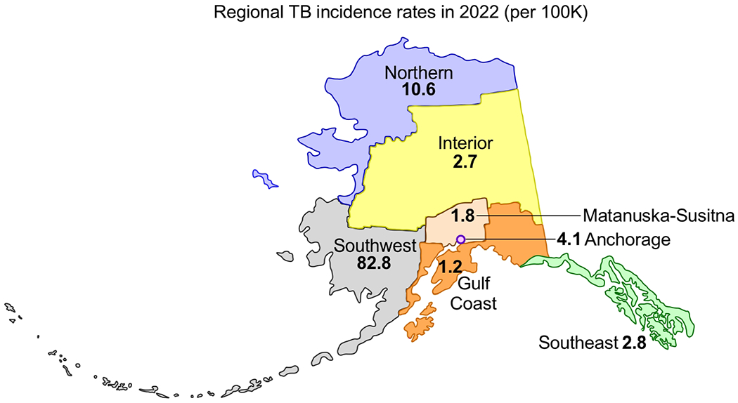
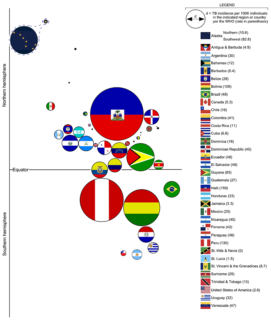
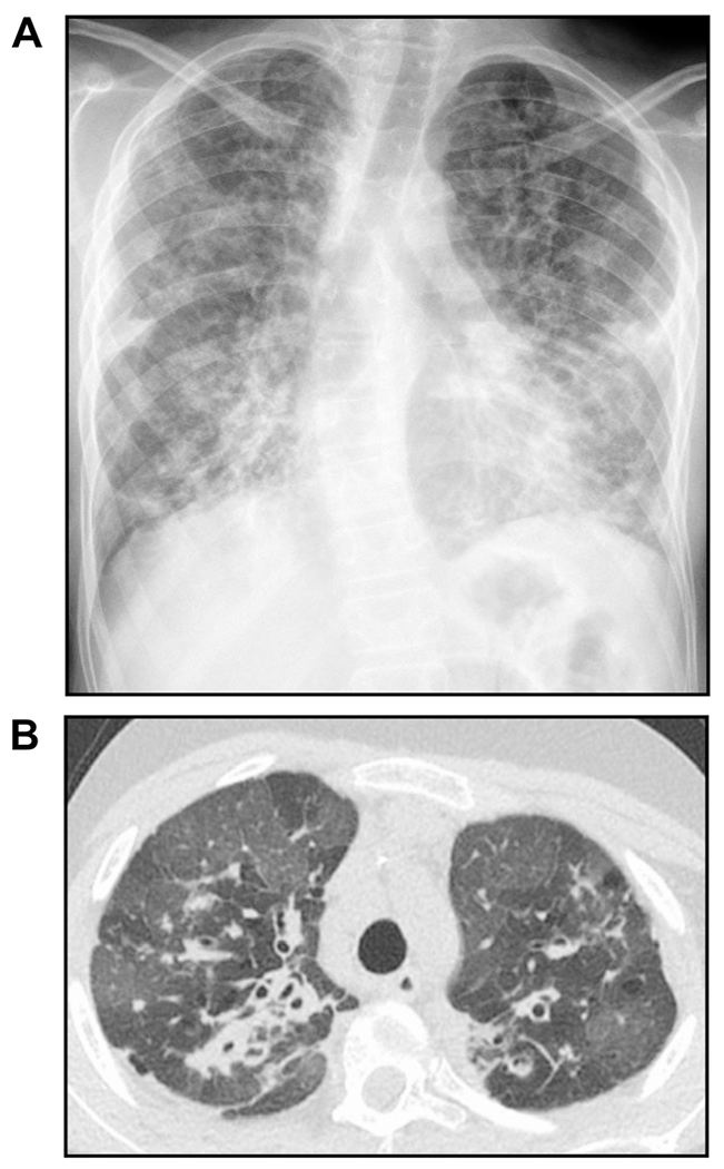
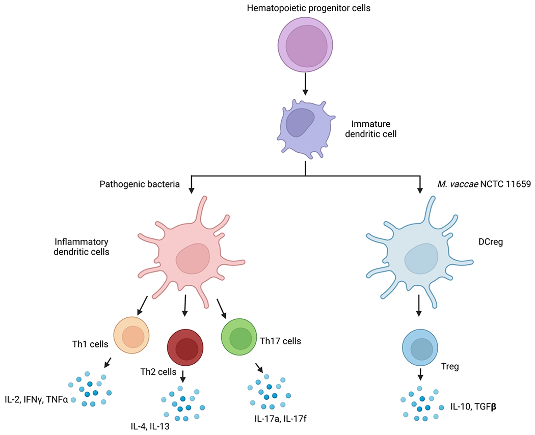
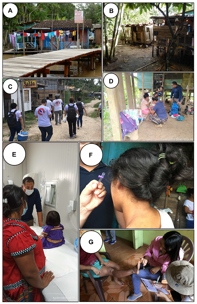
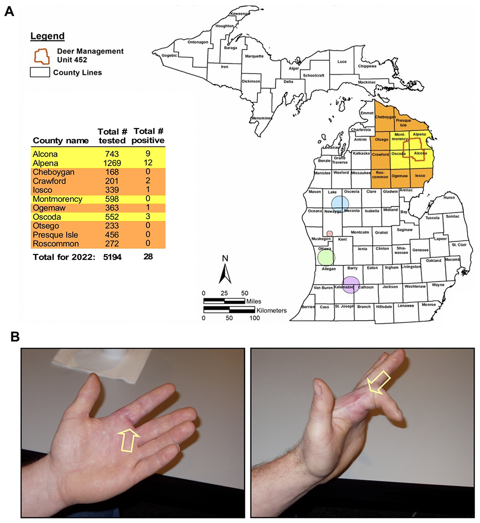
The Many Hosts of Mycobacteria (MHM) meeting series brings together basic scientists, clinicians and veterinarians to promote robust discussion and dissemination of recent advances in our knowledge of numerous mycobacterial diseases, including human and bovine tuberculosis (TB), nontuberculous mycobacteria (NTM) infection, Hansen's disease (leprosy), Buruli ulcer and Johne's disease. The 9th MHM conference (MHM9) was held in July 2022 at The Ohio State University (OSU) and centered around the theme of "Confounders of Mycobacterial Disease." Confounders can and often do drive the transmission of mycobacterial diseases, as well as impact surveillance and treatment outcomes. Various confounders were presented and discussed at MHM9 including those that originate from the host (comorbidities and coinfections) as well as those arising from the environment (e.g., zoonotic exposures), economic inequality (e.g. healthcare disparities), stigma (a confounder of leprosy and TB for millennia), and historical neglect (a confounder in Native American Nations). This conference report summarizes select talks given at MHM9 highlighting recent research advances, as well as talks regarding the historic and ongoing impact of TB and other infectious diseases on Native American Nations, including those in Southwestern Alaska where the regional TB incidence rate is among the highest in the Western hemisphere.


Figures

FIGURE 1.
FIGURE 1.. Tuberculosis among Americans living in Alaska.
The geographical distribution of TB incidence in the state of Alaska for the year 2022, as reported by the Alaska Department of Health for the Northern, Interior, Southwest, Matanuska-Susitna, Gulf Coast and Southeast regions, as well as the capital city Anchorage.
FIGURE 2.
FIGURE 2.. The TB incidence rate of Northern and Southwest Alaska relative to all countries in the Western Hemisphere.
Bubble chart depiction of TB incidence in each country of the Western Hemisphere, per 2021 data reported by the World Health Organization. The diameter of each bubble is directly proportional to TB incidence in the indicated country, each country being represented by its flag. Bubbles are generally organized according to the latitude and longitude coordinates of the corresponding country’s capital city. For comparison purposes, the incidence rates of Northern and Southwest Alaska--the bubble for which are represented by Alaska flag components--are treated separately from all other US states. Note that any country with an incidence of 5/100K or below is depicted by a black dot.
FIGURE 3.
FIGURE 3.. A mausoleum for unknown and unclaimed victims of TB in Sitka, Alaska.
Photo of a World War II ammunition bunker, at Mt. Edgecumbe in Sitka, which was repurposed to be a mausoleum for deceased TB patients in the 1940s and 1950s. No funds were available to transport remains back to their loved ones for half a century. Fifty years later, if relatives could be located, some were transported home for burial. The others remained buried here in Sitka. Image courtesy of Bruce Chandler.
FIGURE 4.
FIGURE 4.. M. bovis infections in wild carnivores.
To survey the prevalence of M. bovis infection and identify their associated cell-mediated immunological biomarkers in wild carnivores or Kruger National Park, South Africa, teams of veterinarians and researchers anesthetize and collect blood from and/or perform endoscopies on (A) spotted hyenas, (B) African wild dogs, and (C-D) lions. Samples are then brought back to laboratory for microbiological and immunological assessments. Images courtesy of Michele Miller.
FIGURE 5.
FIGURE 5.. The outcome of pulmonary infection with Mtb in rabbits.
Depending on the nature of infecting Mtb strain and the infectious inoculum used, rabbits (Oryctolagus cuniculus) can develop the progressive cavitary disease (Active TB; left panel), marked with an elevated bacillary load at 4 weeks that persists until 16 weeks post-infection. These rabbits develop heterogeneous lung granulomas, including necrotic, caseating, and cavitary lesions. The latent Mtb infection (LTBI; mid-panel) model in rabbits is characterized by a transient bacillary growth until 4 weeks that gradually reduces to a complete absence of live bacteria over time (12-24 weeks). These rabbits rarely have any subpleural lesions. Immunosuppression (Kenalog) treatment of rabbits with LTBI leads to the reactivation of active disease (right panel) with elevated bacillary growth in the lungs. The lightweight line is to indicate that immune suppression of rabbits with LTBI results in reactivation of disease. Images courtesy of Selvakumar Subbian.
FIGURE 6.
FIGURE 6.. A case of mycobacteriophage-resistant M. abscessus infection.
(A) Plain chest radiograph and (B) computed tomography of the chest showing advanced bronchiectasis in an adolescent with cystic fibrosis and CFTR genetic mutations not amenable to CFTR modulator. The patient presents for lung transplant evaluation, but that was delayed due to sputum being both AFB stain positive and culture positive for M. abscessus. Standard and investigational therapies were unsuccessful in clearing the positive AFB sputum stain to allow consideration for transplant at that time, so intravenous Mycobacteriophage therapy was instituted that eventually cleared the positive AFB sputum stain. However, after one-year of phage therapy, the patient developed neutralizing antibodies against the phage leading to the return of the AFB sputum stain positivity. Images courtesy of Don Hayes Jr.
FIGURE 7.
FIGURE 7.. Hypothetical model illustrating mechanisms through which “Old Friends”, such as M. vaccae NCTC 11659, impact systemic inflammation and immunoregulation.
Exposure of immature dendritic cells (DC) to M. vaccae NCTC 11659 induces differentiation of regulatory DCs (DCreg) that bias T-cell differentiation away from T helper type 1 (Th1), Th2, and Th17 cells towards induction of regulatory T cells (Treg) that secrete anti-inflammatory cytokines including interleukin 10 (IL-10) and transforming growth factor beta (TGFβ). Abbreviations: DCreg, regulatory dendritic cells; IFNγ, interferon gamma; IL-2, interleukin 2; IL-4, interleukin 4; IL-10, interleukin 10; IL-13, interleukin 13; IL-17a, interleukin 17a; IL-17f, interleukin 17f; NCTC, National Collection of Type Cultures, Th1, T helper type 1; Th2, T helper type 2; Th17, T helper type 17; TNFα, tumor necrosis factor alpha; Treg, regulatory T cells. Figure made with Biorender. Image courtesy of Caelan Wright.
FIGURE 8.
FIGURE 8.. Training the next generation of healthcare workers in Brazil and Panama to recognize signs and symptoms of leprosy.
Scenes from an expedition in Brazil to a fishing village where local healthcare workers were trained to recognize the signs of leprosy. (A) Fishing villages are often accessible only by boat and comprise (B) poor homes in close proximity to frequently flooded river habitats. For this particular trip, (C) a team of leprosy experts and specialists are led by community health agents to the homes of suspected cases. (D) Suspected cases are either examined at their home or (E) in a clinical setting. (F-G) A point-of-care tool being used to improve leprosy diagnosis (Semmes-Weinstein monofilament), seen here being applied to suspected patches of M. leprae infected skin on the (F) cheek and (G) foot. As originally reported in 2021, a Semmes-Weinstein monofilament kit comprises six different monofilaments from the thinnest one, light green (0.07 gm force, like the feeling of a mosquito landing on your skin) to the thickest, pink (>300 gm, the thickness of a pencil lead). The graded force of the six colored monofilaments are used to progressively determine loss of sensation in skin lesions or on the hands or feet by touching the monofilament tip to the skin surface and asking the person if they feel it. If they don’t feel the thickest pink one, then there is total loss of sensation in the skin lesion. Photos courtesy of John Spencer and Carlos Franco-Paredes.
FIGURE 9.
FIGURE 9.. Tuberculosis among white-tailed deer in Michigan.
(A) Deer Management Unit (DMU) 452 is a large area in Michigan, USA, where bovine TB is endemic in white-tailed deer (Odocoileus virginianus). Shown is the location DMU 452 in relation to the counties surrounding it, which are subjected to both active and passive surveillance by the Michigan Department of Natural Resources (DNR), as part of their disease mitigation efforts to prevent spillover to cattle. For active surveillance, hunters voluntarily submit the heads of deer for examination; for passive surveillance, hunters may submit deer carcasses with TB chest lesions from anywhere in the state. In yellow and orange are counties surrounding DMU 452, where surveillance efforts are most focused, as well as distal testing sites (circled) to surveille bovine TB movement south (the Great Lakes prevent bovine TB movement north). The table inset lists for each county surrounding DMU 452 the total number of deer tested and total positive for M. bovis in 2022, as originally reported by the Michigan DNR. (B) An M. bovis-infected hand, the result of a zoonotic transmission event which occurred while preparing an M. bovis-infected deer head for taxidermy. Images courtesy of James Sunstrum.
FIGURE 10.
FIGURE 10.. Progression of post-primary pulmonary TB lesions is associated with macrophage polarization.
Post-primary pulmonary TB lesions in B6.Sst1S mice 14 weeks after footpad infection with 106 of virulent Mtb Erdman. Shown are (A) a controlling paucibacillary lesion, and (B) a non-controlling multibacillary lesion, with stains corresponding to T lymphocytes (CD3+, red), B lymphocytes (CD19+, green), and macrophages (Iba1+, blue) expressing nitric oxide synthase (iNOS+, magenta) or arginase (Arg1+, yellow). Scale bare = 50 microns. Acid fast staining on serial sections was used to classify TB lesions: controlling lesions were paucibacillary and Mtb was intracellular with 1-2 bacilli per infected macrophage. The non-controlling lesions were multibacillary with clusters of intracellular Mtb and areas of micronecrosis containing extracellular bacteria. Multiplexed fluorescent immunohistochemistry and quantification using HALO software were performed as described in Rosenbloom et al . Images courtesy of Igor Kramnik and Nicholas Crossland.

References

    1. Schildknecht KR, Pratt RH, Feng PI, Price SF, Self JL. Tuberculosis - United States, 2022. MMWR Morb Mortal Wkly Rep 2023;72:297–303. doi: 10.15585/mmwr.mm7212a1. - DOI - PMC - PubMed
    1. Ehrenpreis JE, Ehrenpreis ED. A Historical Perspective of Healthcare Disparity and Infectious Disease in the Native American Population. Am J Med Sci 2022;363:288–294. doi: 10.1016/j.amjms.2022.01.005. - DOI - PMC - PubMed
    1. Bloss E, Holtz TH, Jereb J, Redd JT, Podewils LJ, Cheek JE, McCray E. Tuberculosis in indigenous peoples in the U.S., 2003-2008. Public Health Rep 2011;126:677–689. doi: 10.1177/003335491112600510. - DOI - PMC - PubMed
    1. Daniel TM. The history of tuberculosis. Respir Med 2006;100:1862–1870. doi: 10.1016/j.rmed.2006.08.006. - DOI - PubMed
    1. Gagneux S. Host-pathogen coevolution in human tuberculosis. Philos Trans R Soc Lond B Biol Sci 2012;367:850–859. doi: 10.1098/rstb.2011.0316. - DOI - PMC - PubMed

Publication types

LinkOut - more resources